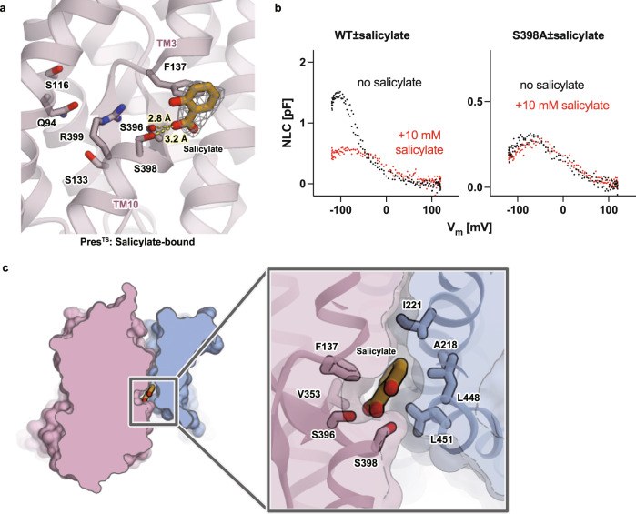
Fig. 3. Salicylate binding.
a A close-up view of the central anion-binding pocket of PresTS with a bound salicylate. Salicylate and some important residues are shown in stick representations. The cryo-EM density of salicylate is shown as a gray mesh. Predicted short-distance interactions are indicated by yellow dotted lines with the distance. b The effect of salicylate on the NLC of WT (left) and S398A-HgPres (right). NLC was measured from the same cells before (black) and after (red) the application of 10 mM salicylate in the bath solution. The total numbers of recordings were seven for both WT- and S398A-HgPres. Source data are provided as a Source Data file. c Salicylate binding pocket. A close-up view of the salicylate binding pocket between the core and the gate domains of PresTS (left panel). Salicylate is sandwiched between the core and the gate domains of PresTS (right panel).

