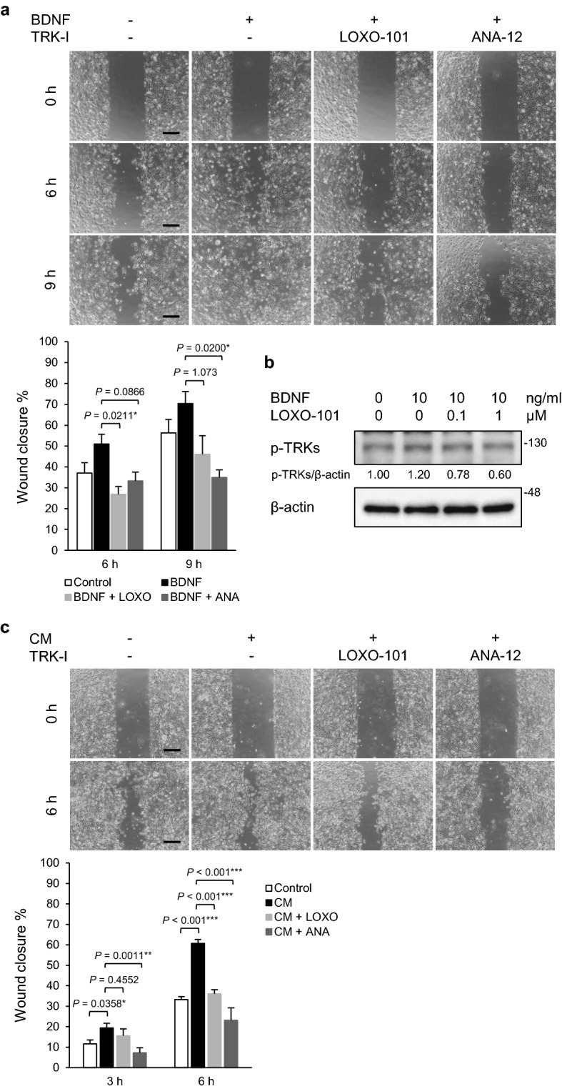
Figure 4.

Suppression of BDNF-induced cell migration of PGC cells by TRK inhibitors. (a) Wound healing assay. After serum starvation (0.5% FBS for 24 h), PGC cells were pre-treated with LOXO-101 (a pan-TRK inhibitor, 100 nM) or ANA-12 (a TRKB specific inhibitor, 10 μM) for 1 h, and a wound healing assay was performed. The migrating cells were imaged and cell migration was estimated as wound closure percentage at the indicated time points (0, 6, and 9 h). Histograms depict the percentages of wound closure as the means ± SEM of triplicate wells from three independent experiments. Scale bars, 100 μm. *P < 0.05 (Tukey’s test). (b) Phosphorylation of TRKB in PGC cells was determined by WB analysis. After serum starvation (0.5% FBS for 24 h), PGC cells were pre-treated with LOXO-101 (100 nM) for 1 h and subsequently cultured in the presence or absence of BDNF (10 ng/mL) for 30 min. The relative amounts of p-TRKs are indicated as the ratio of each protein band relative to the loading control, β-actin. (c) Wound healing assay with CM from PGC/CAF co-cultures. Data shown are means ± SEM of quintuple wells and are representative of three independent experiments. TRK-I, TRK inhibitor. ***P < 0.001, **P < 0.01, and *P < 0.05 (Tukey’s test).
