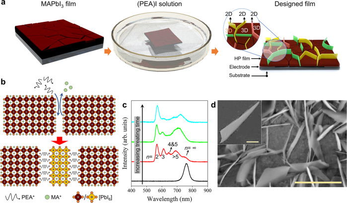
Fig. 1. Isolating 3D HP grains by 2D HP platelets.
a Schematic diagram of the experimental fabrication process of the GIHP film. b Schematic illustration of the cation exchange-induced growth of 2D HP at the boundaries of 3D HP grains. c Treating time-dependent PL spectra of the film. d The SEM micrograph of the obtained sample. The scale bar in is 5 μm. Inset: Zoomed view of the 2D HP platelet inserted in the grain boundary, and the scale bar is 500 nm.

