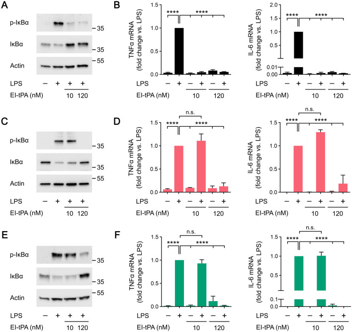
Figure 4.
The anti-inflammatory activity of EI-tPA is facilitated by membrane-anchored PrPC and LRP1. (A) BMDMs isolated from WT mice were treated for 1 h with LPS (0.1 μg/mL), LPS plus 10 nM El-tPA, LPS plus 120 nM El-tPA, or vehicle. Cell extracts were subjected to immunoblot analysis to detect phospho-IκBα, total IκBα, and β-actin. (B) WT BMDMs were treated with LPS (0.1 μg/mL), LPS plus 10 nM El-tPA, LPS plus 120 nM El-tPA, or vehicle for 6 h. RT-qPCR was performed to compare mRNA levels for TNFα and IL-6 (n = 4–6). (C) PrPC-deficient BMDMs isolated from Prnp−/− mice were treated for 1 h with LPS (0.1 μg/mL), LPS plus 10 nM El-tPA, LPS plus 120 nM El-tPA, or vehicle. Cell extracts were subjected to immunoblot analysis to detect phospho-IκBα, total IκBα, and β-actin. (D) PrPC-deficient BMDMs were treated with LPS (0.1 μg/mL), LPS plus 10 nM El-tPA, LPS plus 120 nM El-tPA, or vehicle for 6 h. RT-qPCR was performed to compare mRNA levels for TNFα and IL-6 (n = 4–5). (E) LRP1-deficient BMDMs isolated from mLrp1−/− mice were treated for 1 h with LPS (0.1 μg/mL), LPS plus 10 nM El-tPA, LPS plus 120 nM El-tPA, or vehicle. Cell extracts were subjected to immunoblot analysis to detect phospho-IκBα, total IκBα, and β-actin. (F) LRP1-deficient BMDMs were treated with LPS (0.1 μg/mL), LPS plus 10 nM El-tPA, LPS plus 120 nM El-tPA, or vehicle for 6 h. RT-qPCR was performed to compare mRNA levels for TNFα and IL-6 (n = 4–5). Data are expressed as the mean ± SEM (One-way ANOVA; ****P < 0.0001, n.s.: not statistically significant).

