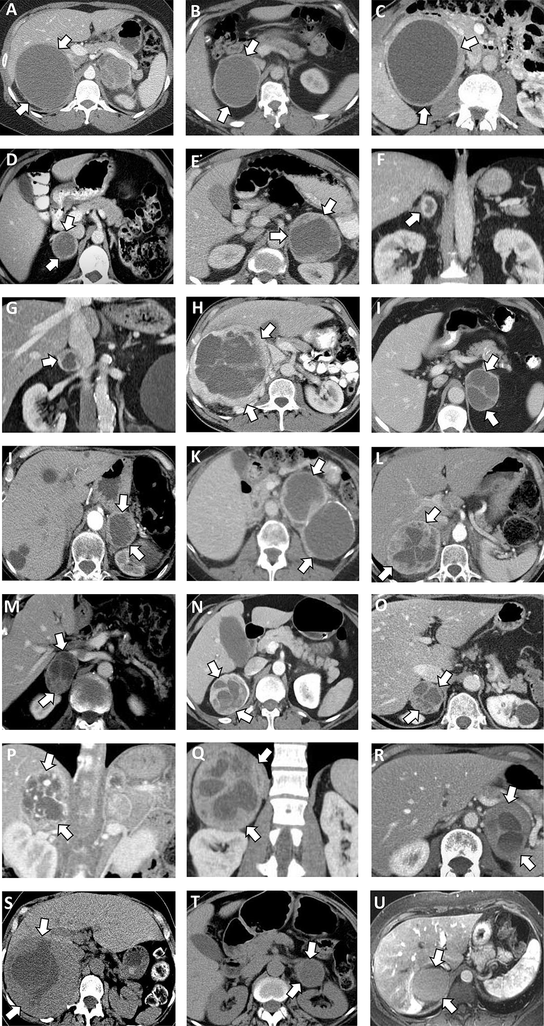
Figure 1. Computed tomography (CT) and magnetic resonance imaging (MRI) imaging of 21 patients with cystic pheochromocytomas.

(A) Right 11.2 cm pheochromocytoma, cystic component 75%, on contrast enhanced CT.
(B) Right 9.1 cm pheochromocytoma, cystic component 77% on contrast enhanced CT.
(C) Right 11.7 cm pheochromocytoma, cystic component 68% on contrast enhanced CT.
(D) Right 4.1 cm pheochromocytoma, cystic component 26% on contrast enhanced CT.
(E) Left 8.2 cm pheochromocytoma, cystic component 50% on contrast enhanced CT.
(F) Left 2.9 cm pheochromocytoma, cystic component 15% on contrast enhanced CT.
(G) Right 3.8 cm pheochromocytoma, cystic component 24% on contrast enhanced CT.
(H) Right 18.9 cm pheochromocytoma, cystic component 40% on contrast enhanced CT.
(I) Left 6.4 cm pheochromocytoma, cystic component 53% on contrast enhanced CT.
(J) Left, 5.0 cm pheochromocytoma, cystic component 50% on contrast enhanced CT.
(K) Left 12.0 cm pheochromocytoma cystic component 64%, peripheral calcification on contrast enhanced CT.
(L) Right 8.2 cm pheochromocytoma, cystic component 28% on contrast enhanced CT.
(M) Right 5.5 cm pheochromocytoma, cystic component 44% on contrast enhanced CT.
(N) Right 6.5 cm pheochromocytoma, cystic component 29% on contrast enhanced CT.
(O) Right 4.4 cm pheochromocytoma, cystic component 24% on contrast enhanced CT.
(P) Right 5.0 cm pheochromocytoma, cystic component 24% on contrast enhanced CT.
(Q) Right 13.5 cm pheochromocytoma, cystic component 23% on contrast enhanced CT.
(R) Left 6.3 cm pheochromocytoma, cystic component 25% on contrast enhanced CT.
(S) Right 14.9 cm pheochromocytoma, cystic component 22% on unenhanced CT.
(T) Left 5.7 cm pheochromocytoma, cystic component 56% on unenhanced CT.
(U) Right 6.3 cm pheochromocytoma, cystic component 70% on contrast enhanced MRI.
