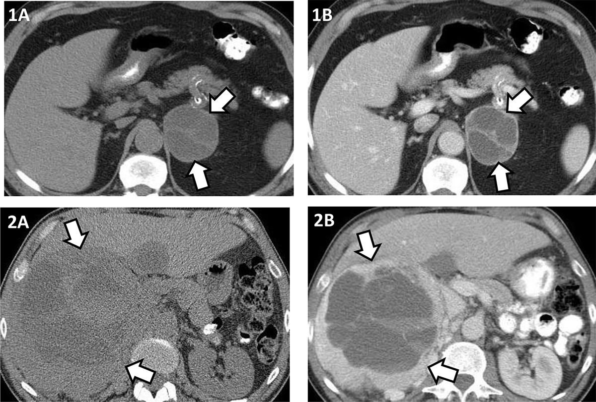
Figure 2: Illustrating cystic pheochromocytoma appearance on unenhanced and contrast enhanced computed tomography (CT).

1A and 1B – Left 6.4 cm pheochromocytoma, cystic component 14 HU, solid component 34 HU on unenhanced CT (1A); cystic component 20 HU, solid component 90 HU, rim and septal enhancement on contrast enhanced CT (1B).
2A and 2B –Right 18.9 cm pheochromocytoma, cystic component 18 HU, solid component 35 HU on unenhanced CT (2A); cystic component 21 HU, solid component 132 HU, rim enhancement on contrast enhanced CT (2B).
