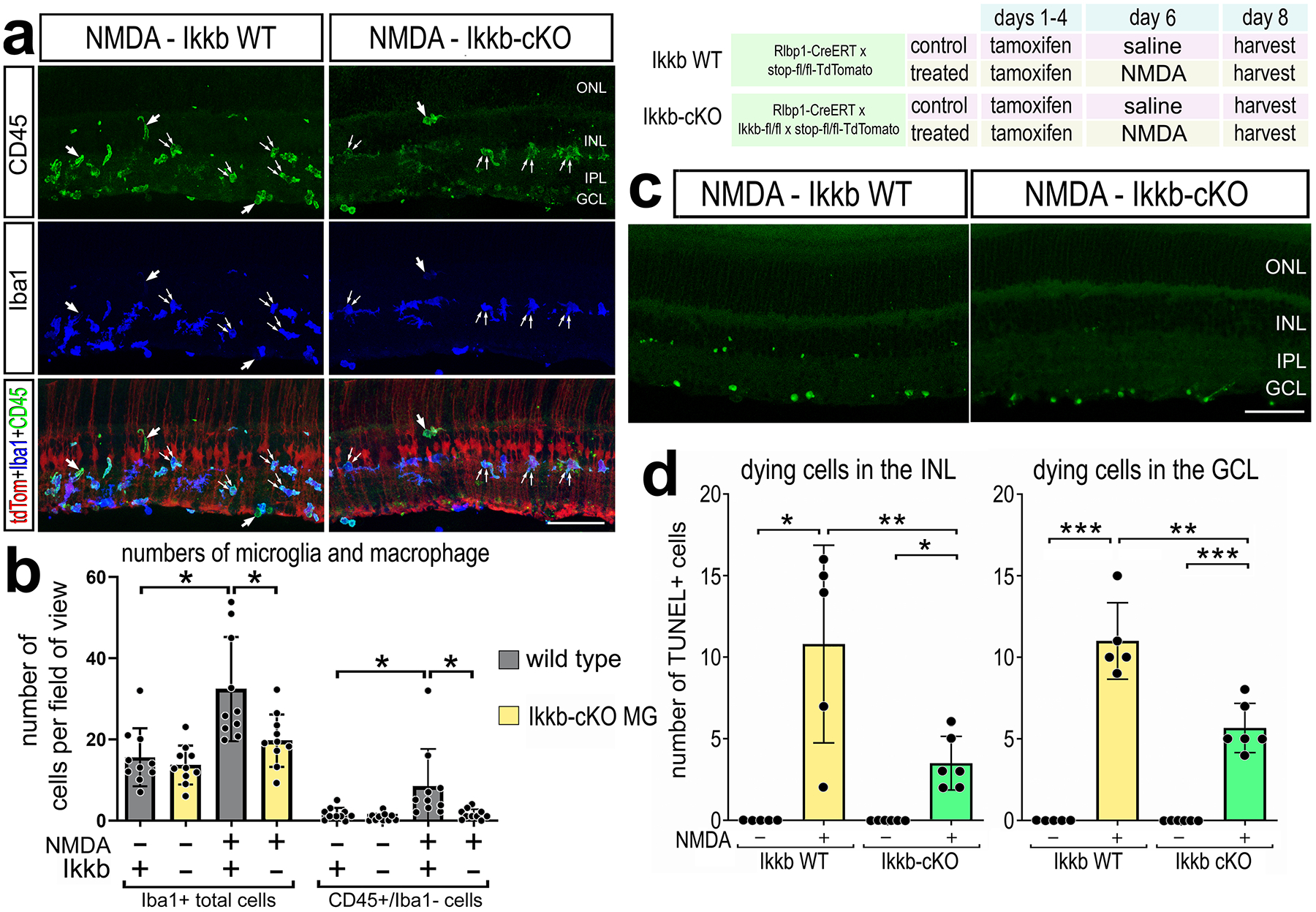
Figure 3: Conditional knockout of NFkB in Müller glia impairs immune cell responses after damage.

Rlbp1-creERT and Rlbp1-creERT:Ikkbfl/fl mice were injected IP with tamoxifen 1x daily for 4 consecutive days. Left eyes were injected with saline and right eyes were injected with NMDA on D6, and retinas were harvested on D8. (a) Retinal sections were labeled for CD45 (green), Iba1 (blue), and TdTomato (red). Single arrows in a represent CD45+/Iba1− cells; double arrows indicate CD45+/Iba1+ cells. The histogram illustrates the mean number (±SD and individual data points) of Iba1+ or CD45+/Iba1− cells and significance of difference (*p<0.05) was determined by a Kruskal-Wallis test (b). TUNEL assays were performed on retinal sections to identify dying cells. Histogram illustrate the mean number (±SD and individual data points) of TUNEL+ cells in the INL and GCL and significance of difference (*p<0.05, **p<0.01) was determined by paired t-test (d). The calibration bars in panels a,c represent 50 μm. Abbreviations: ONL – outer nuclear layer, INL – inner nuclear layer, IPL – inner plexiform layer, GCL – ganglion cell layer.
