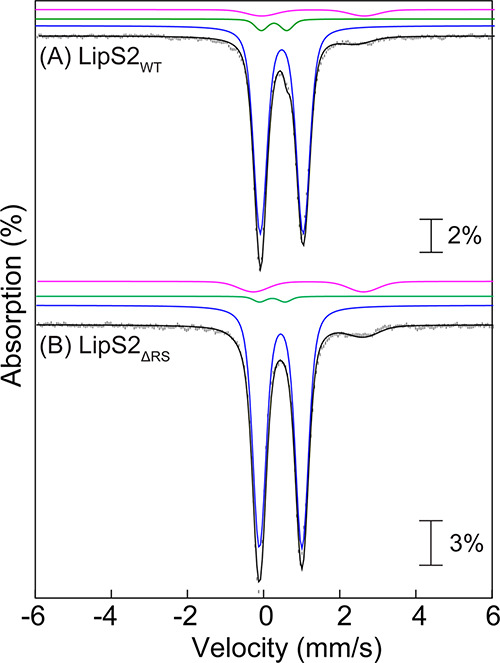
Figure 5.

Mössbauer spectra of reconstituted (A) LipS2WT and (B) LipS2ΔRS collected at 4.2 K (vertical bars) in the absence of any external applied magnetic field. The black lines represent the overall simulated spectra, while the individual contributions from the [Fe4S4]2+ (90% of total intensity in both LipS2WT and LipS2ΔRS), [Fe2S2]2+ (5 and 2% of total intensities in LipS2WT and LipS2ΔRS, respectively) clusters, and N/O-coordinated high-spin FeII (5 and 8% of total intensities in LipS2WT and LipS2ΔRS, respectively) are shown by the blue, green, and pink lines, respectively.
