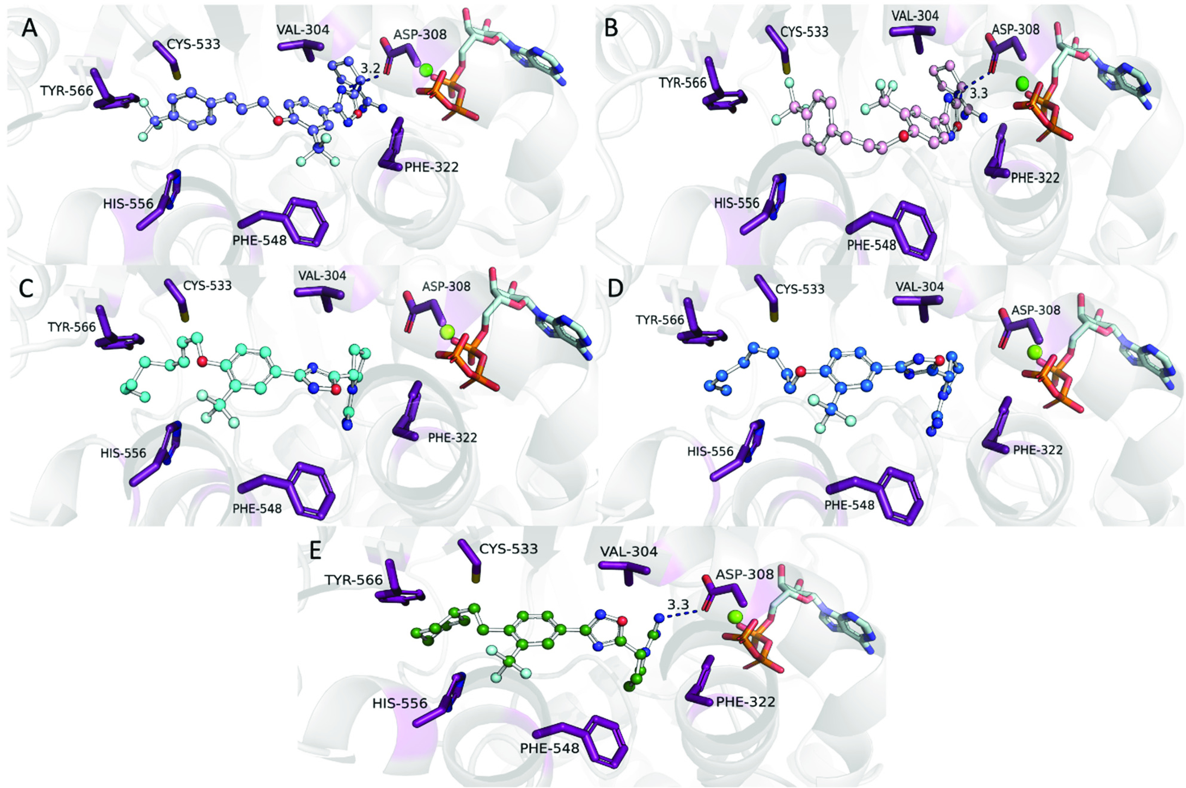
Figure 4.

Molecular docking of top compounds indicates positioning of inhibitors in the back of the binding pocket influence binding potency. Molecular docking of 23d (blue, A) and 23e (pink, B) in an hSphK2 homology model position the ligand toward the top of the binding cavity near ATP. The hSphK2 homology model is based on the template of hSphK1 with sphingosine cocrystallized (PDB ID: 3VZB). Analysis of alkene bond geometry in alkyl tail analogues 26d (cyan, C) and 26a (blue, D) through molecular docking in an hSphK2 homology model show utilization of the side pocket near Phe548. Compound 14c (green, E) utilizes the side pocket near Phe548 to position the trifluoromethyl group and participates in electrostatic interactions with Asp308. Hydrogen bonds are shown in blue dashed lines.
