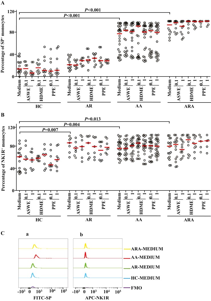
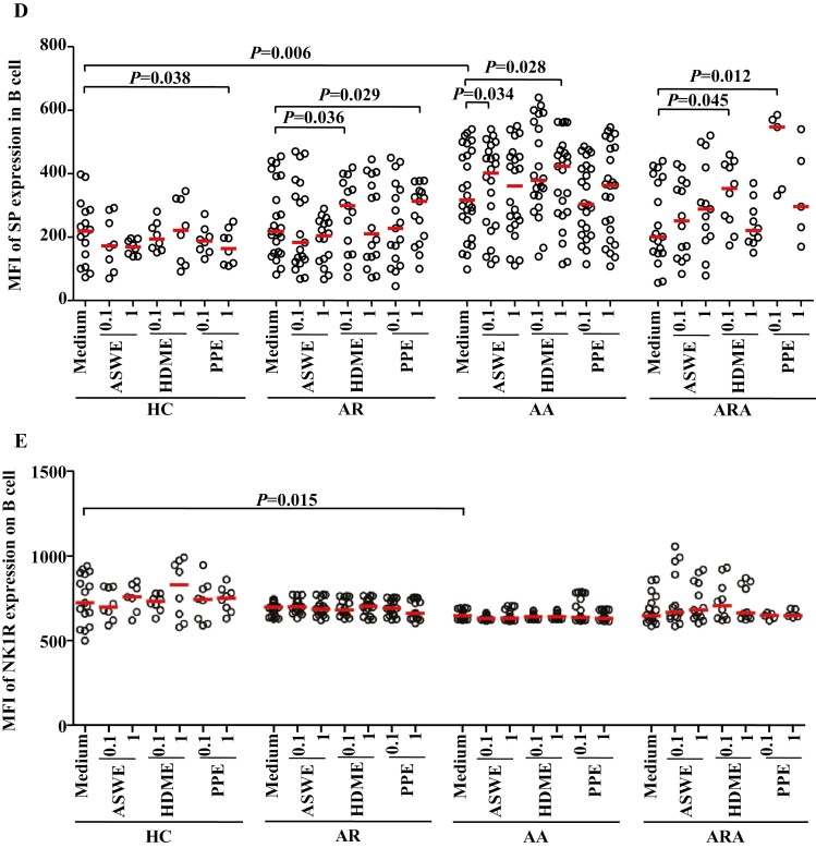
Figure 2:
Flow cytometry analysis of expressions of SP and NK1R in human peripheral blood monocyte (CD14+ cells). (A) and (B) represented the percentages of SP+ and NK1R+ monocytes in the patients with allergic rhinitis (AR), allergic asthma (AA), AR combined with AA (ARA), and healthy control (HC) subjects, respectively. (Ca) and (Cb) showed representative flow cytometric figures of mean fluorescent intensity (MFI) of SP and NK1R expressions in the monocytes of AR, AA, ARA, and HC subjects. (D) and (E) represented the MFI of SP and NK1R in the monocytes of AR, AA, ARA, and HC subjects. Cells were stimulated with or without house dust mite extract (HDME), Artemisia sieversiana wild allergen extract (ASWE), or Platanus pollen allergen extract (PPE), all at the concentrations of 0.1 and 1 µg/ml for 1 h at 37°C. Each symbol represents the value of one subject. The median value is indicated by a horizontal line. P < 0.05 was taken as statistically significant. FMO: fluorescence minus one.


