Figure 1:
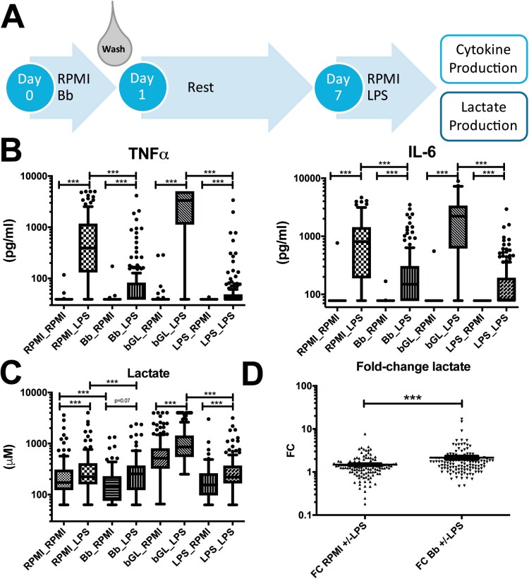
Inflammatory response of primary monocytes after exposure to B. burgdorferi. (A) Schematic representation of the trained immunity model. Primary monocytes are stimulated for 24 hour with medium control (RPMI), B. burgdorferi (Bb, MOI = 1), β-glucan (bGL, 2µL) or LPS (10 ng/ml). After 24 hour, the stimuli are washed away and cells are rested for six days, after which cells are restimulated with RPMI or LPS (10 ng/ml). After 24 hour, cell-free supernatants are collected for analysis of lactate- and cytokine production. (B) TNFα and IL-6 production in cell-free supernatants from monocytes of 151 healthy volunteers subjected to the trained immunity protocol described above. (C) Lactate concentration (µM) in cell-culture supernatant from primary monocytes of 120 healthy volunteers subjected to the trained immunity protocol described above. (D) Fold-change (FC) in lactate production after restimulation with LPS versus mock-treatment in primary monocytes previously exposed to B. burgdorferi or RPMI (n = 120). *** P < 0.0001 test with Wilcoxon matched-pairs signed rank test in panels B-D.

