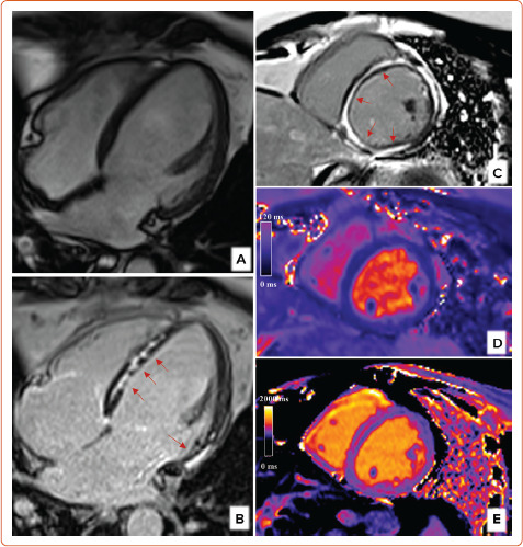
Figure 1: Cardiac Magnetic Resonance in Dilated Cardiomyopathy.

In this patient with dilated cardiomyopathy, cardiac magnetic resonance provides accurate assessment of left ventricular volumes and function by balanced steady state free precession sequences (A). Cardiac magnetic resonance also identifies the presence of mid-wall late gadolinium enhancement in mid-to basal septum and basal lateral wall (B, red arrows). The short axis at mid-ventricular level reveals almost circumferential mid-wall late gadolinium enhancement (C, red arrows); T2 values within the normal ranges exclude the presence of myocardial oedema (D). Native T1 values were instead globally increased (E).
