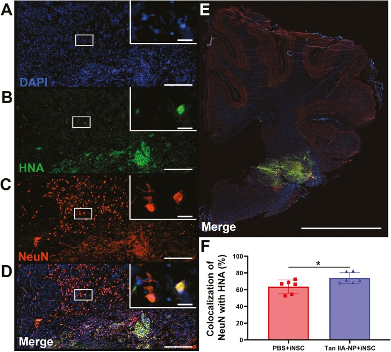
Figure 2.
Tan IIA-NP treatment enhanced iNSC neuronal differentiation. Immunohistochemistry analysis showed human HNA + cells colocalized with the mature neuron protein NeuN at the lesion site 12 weeks post-transplantation (A-D; scale bar = 300 µm; white box; scale bar = 10 µm). Quantitative analysis of PBS + iNSC and Tan IIA-NP + iNSC ipsilateral hemisphere scans (E; scale bar = 10 mm) revealed Tan IIA-NPs increased the number of HNA+/NeuN + cells at 12 weeks post-transplantation (73.96 ± 5.86% vs. 63.61 ± 7.39%, respectively; F). Data are expressed as mean ± SD. * indicates a significant (P < .05) difference between treatment groups. PBS + PBS data n = 6, PBS + iNSC data n = 6, and Tan IIA-NP + iNSC data n = 6.

