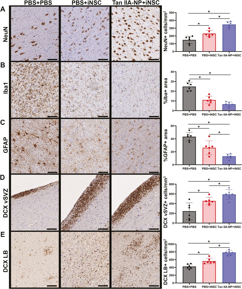
Figure 4.
Tan IIA-NPs enhanced iNSC induced neuroprotective and regenerative activity. Tan IIA-NP + iNSC pigs showed an increased number of NeuN + neurons at 12 weeks post-transplantation relative to PBS + iNSC and PBS + PBS pigs (346.96 ± 34.45 cells/mm2 vs. 230.69 ± 37.81 cells/mm2 vs. 149.29 ± 43.57 cells/mm2, respectively; A). Tan IIA-NP + iNSC pigs also showed decreased numbers of Iba1 + immune cells relative to PBS + iNSC and PBS + PBS pigs (6.61 ± 2.21% vs. 10.98 ± 3.54% vs. 24.78 ± 4.05%, respectively; B). Similarly, Tan IIA-NP + iNSC pigs also showed fewer GFAP + reactive astrocytes relative to PBS + iNSC and PBS + PBS pigs (13.00 ± 3.36% vs. 26.20 ± 9.81% vs. 43.28 ± 6.02%, respectively; C). Tan IIA-NP + iNSC pigs demonstrated increased numbers of DCX + neuroblasts in the ventricular lining of the subventricular zone (vSVZ) relative to PBS + iNSC and PBS + PBS pigs (589.14 ± 90.49 cells/mm2 vs. 454.33 ± 68.36 cells/mm2 vs. 245.04 ± 156.11 cells/mm2, respectively; D) and at the lesion border (LB; 783.60 ± 56.69 cells/mm2 vs. 562.41 ± 65.01 cells/mm2 vs. 424.77 ± 54.70 cells/mm2, respectively; E). Scale bars 500 µm. Data are expressed as mean ± SD. PBS + PBS data n = 6, PBS + iNSC data n = 6, and Tan IIA-NP + iNSC data n = 6.

