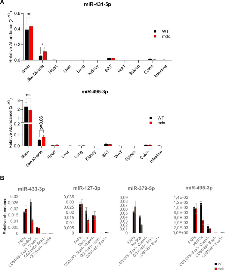
Figure S1. DD-miRNAs expression level in different organs, tissues and skeletal muscle cellular subpopulations.
(A) Comparison of expression levels of 2 representatives DD-miRNAs (miR-431-5p and miR-495-3p) between different organs from 5-wk-old wild-type and mdx mice (n = 3–4). (B) Comparison of expression levels of four representative DD-miRNAs between different FACS-sorted cell populations from 5-wk-old mdx mice and its healthy controls (n = 3). Muscle mono-nucleated cells (MMNC) fractions were FACS-sorted by using CD31, CD45, Sca1, and Vcam1 markers from hind limb muscles of mdx and control mice. We quantified the expression of four representative DD-miRNAs (n = 3) in five subpopulations that are typically found in skeletal muscle, which are the hematopoietic cells (CD45+), endothelial cells (CD31+), fibro-adipocyte progenitors (FAPs) (CD31−/CD45−/Sca1+), satellite cells (CD31−/CD45−/Sca1−/Vcam1+), and cells that are negative for all four markers (CD45−/CD31−/Sca1−/Vcam1−). Expression of DD-miRNAs was detected in FAPs, satellite cells, and CD31/CD45/Sca1/Vcam1 negative cellular fraction. Comparison between the mdx and WT cells showed that DD-miRNAs were expressed to similar levels in the FAPs and in the negative cell population. In contrast, a lower DD-miRNAs expression was detected in satellite cells derived from the mdx mouse compared with control, which is consistent with the activated state of satellite cells of the mdx mouse, and with the fact that DD-miRNAs are known to be down-regulated in activated satellite cells (Castel et al, 2018). Data are presented as mean ± SEM. *P < 0.05; **P < 0.01; ***P < 0.001.

