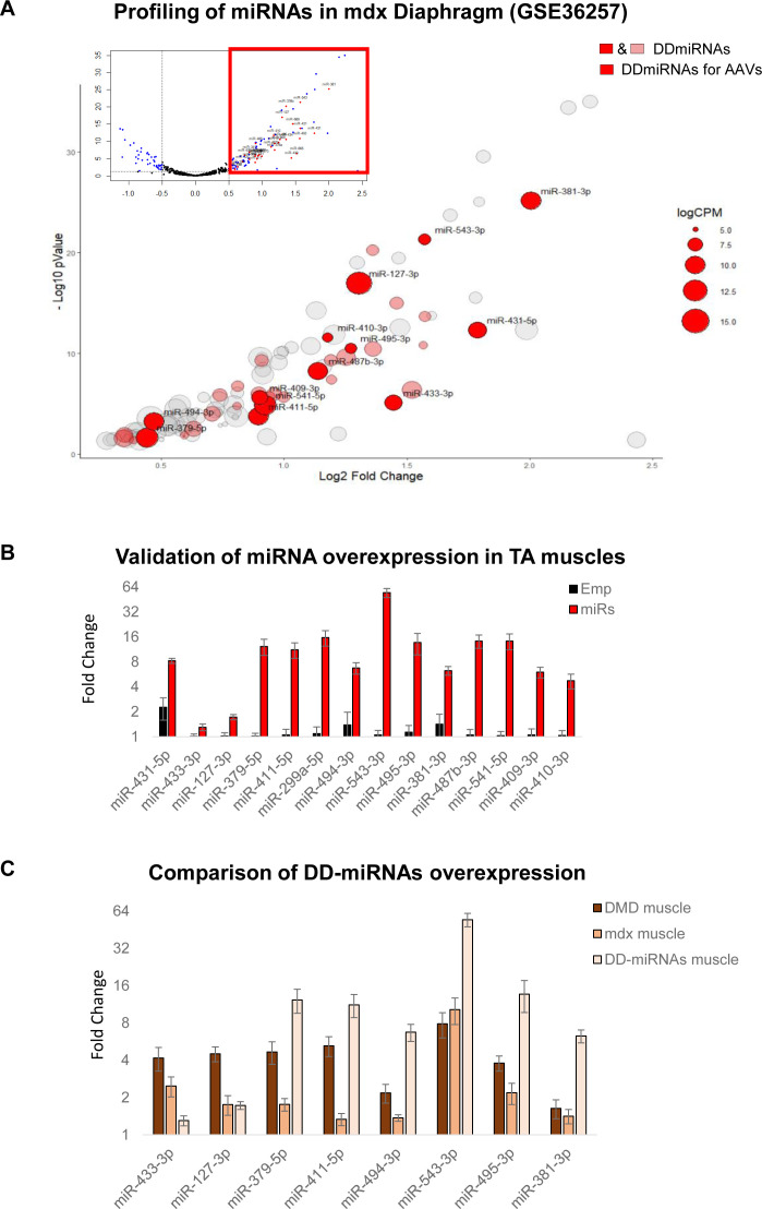
Figure S2. Selection and in vivo overexpression of 14 DD-miRNAs in skeletal muscle.
(A) Volcano plot of miRNA profiling of the diaphragm of 8-wk-old mdx and control mice (upper part), and a zoom-in of significantly up-regulated miRNAs in mdx muscle (lower part) (data were taken and reanalyzed from GEO: GSE36257 [Roberts et al, 2012]). Expression levels are presented by the size of the circle. DD-miRNAs are colored (pink or red). MiRNAs in red were selected for the overexpression by AAV vectors in the present study. (B) Validation of DD-miRNAs overexpression in the treated mice. TA muscles were analyzed 1 mo after injection with AAV-DD-miRNAs (miRs) compared with AAV-Empty control (Emp) (n = 6). (C) Comparison of DD-miRNAs overexpression in different systems: muscle biopsies from Duchenne muscular dystrophy patients compared with healthy controls (Fig 1A), diaphragm muscle of mdx mice compared with C57Bl10 control (Fig 1B), and DD-miRNAs overexpression in the TA muscle of a healthy mouse by AAV vectors. Data in (B, C) are presented as mean ± SEM.

