Fig. 5.

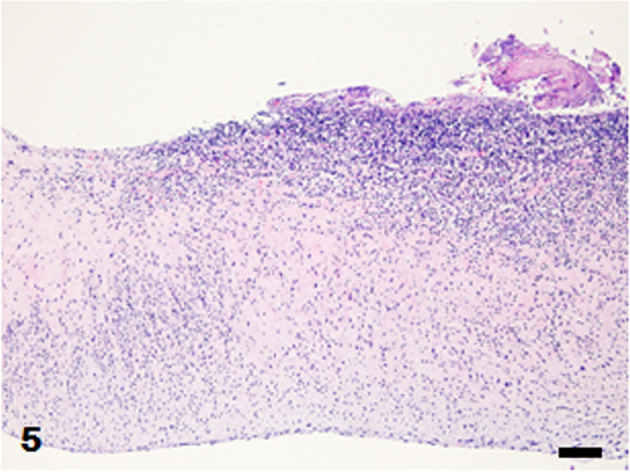
Mitral valve. Numerous inflammatory cells infiltrate the valve wall. Fibrinous exudates with cocci are attached to the valve. Hematoxylin and eosin (HE) stain. Bar=100 µm.

Mitral valve. Numerous inflammatory cells infiltrate the valve wall. Fibrinous exudates with cocci are attached to the valve. Hematoxylin and eosin (HE) stain. Bar=100 µm.