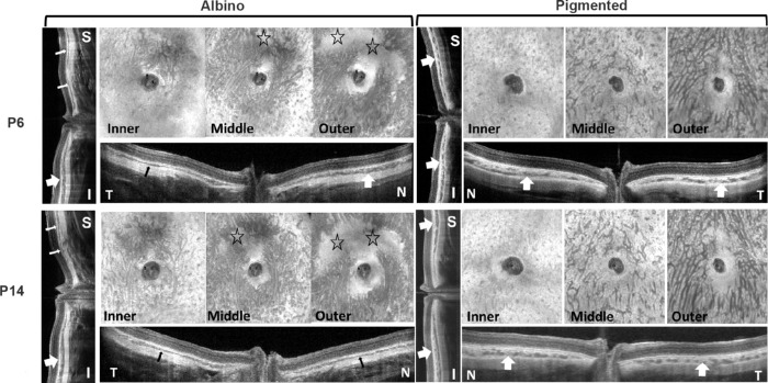
Figure 6.
En face and cross-sectional images from representative pigmented and albino GP eyes at P6 (upper images) and P14 (lower images) showing the development of vascular layers of the choroid. S, superior; I, inferior; T, temporal; N, nasal. Stars denote underdeveloped choroidal areas in the albino eye. All images were obtained by SS-OCT. Thin arrows indicate thin choroid, and thick arrows indicate thick choroid—showing the unevenness of ChT in albino GPs and more even ChT in pigmented GPs in different quadrants.

