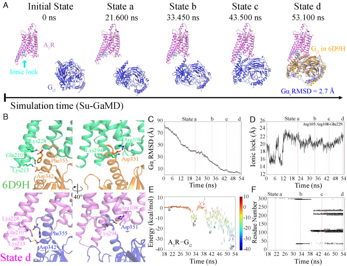
Fig. 3.
(A) The landscape of the A1R−Gi2 recognition pathway. The relative position of Gi2 after global alignment of A1R (A1R is shown in violet, and Gi2 is shown in blue) to that of the 6D9H structure (Gi2 is shown in orange) is shown in state d. (B) The same key molecular interactions in the 6D9H structure (A1R and Gi2 are shown in green and orange, respectively) and state d (A1R and Gi2 are shown in violet and blue, respectively). Time-dependent (C) Gαi rmsd, (D) N–O distance between the guanidinium of Arg1053.50/Arg1083.53 and the carboxyl of Glu2296.30, (E) the binding free energy landscape for A1R−Gi2, and (F) A1R−Gi2 contact residues during the recognition process.

