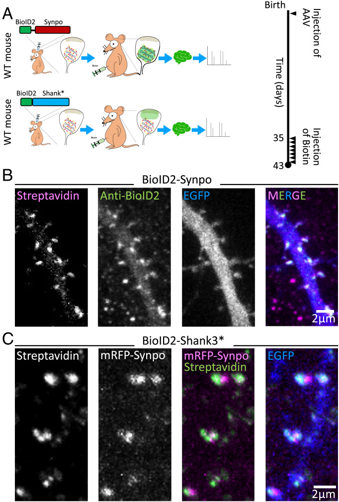
Fig. 2.
Identification of SA-associated proteins by in vivo proximity labeling. (A) Schematic representation of in vivo proximity labeling of protein neighbors of synaptopodin (iBioID). To target the biotinylating enzyme to the SA, AAV2/9 viruses containing BioID2-synaptopodin were injected in the cortex of neonatal mice. BioID2 fused to a fragment of Shank3 (BioID-Shank3*) was used as a control to localize the biotinylating enzyme to a different region of the spine (i.e., the neighborhood of the PSD). Biotin was administered intraperitoneally starting 5 wk after birth for 7 consecutive days. On day 8, the mouse was euthanized, and biotinylated proteins were isolated from their brain. WT: wild type. (B) Cultured hippocampal neurons transfected with BioID2-synaptopodin and EGFP were stained with Alexa647-streptavidin and anti-BioID2 antibody to confirm the specificity of biotinylation in spines. (C) A cultured hippocampal neuron expressing mRFP-synaptopodin, BioID2-Shank3*, and EGFP was labeled with streptavidin to examine the specificity of biotinylation as well as the differential localization of synaptopodin and of the control protein Shank3* in spines.

