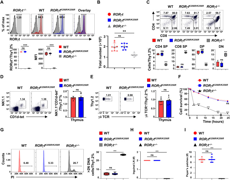
Fig. 3. RORγt-K256R mutation does not affect RORγt-dependent development of thymocytes and lymph nodes.
(A) Flow cytometric analysis of RORγt in thymocytes obtained from indicated mice (n = 5 to 8). MFI, mean fluorescence intensity. (B) Total thymocyte numbers of the indicated mice (n = 5 to 8). The graph shows mean ± SD. (C) Flow cytometric analysis of CD4 and CD8 on thymocytes from indicated mice (n = 4 per genotype). (D) Representative flow cytometric analysis of NKT cells (n = 4) from the thymus of the indicated mice. (E) Representative flow cytometric analysis of γδ T cells (n = 4) from the thymus of the indicated mice. (F) Flow cytometric analysis of the survival of DP thymocytes cultured in vitro for the indicated time (n = 4 per genotype). (G) Representative flow cytometric analysis of DNA content of indicated thymocytes stained by propidium iodide (PI) (n = 4 to 6 per genotype). (H and I) Number of inguinal lymph nodes (LN) (H) and Peyer’s patches (I) in the indicated mice (n = 9 to 12 per genotype). **P < 0.01; ***P < 0.001 (two-tailed t test). (A, C, E, F, and G) Box plots or scatter plots show median (central line), maximum, minimum (box ends), and outliers (extended lines).

