Figure 1.
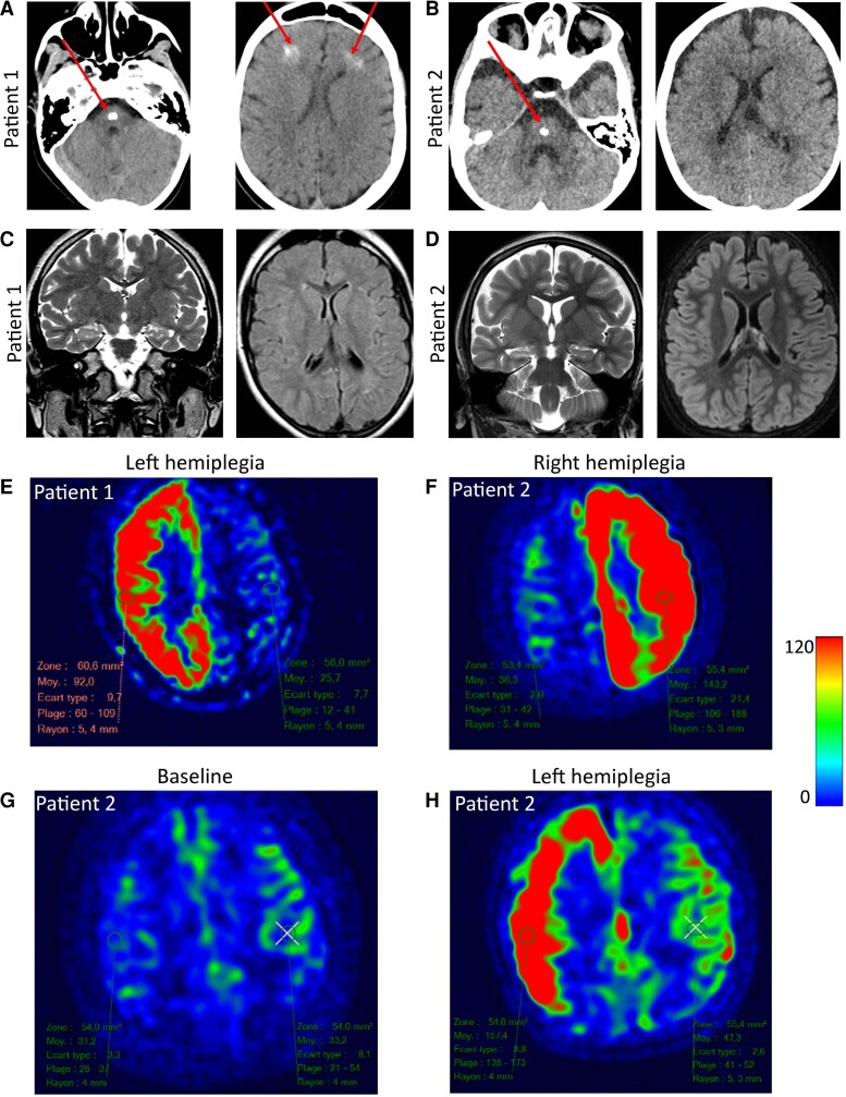
Neuroimaging of patients with hemiplegia. (A) Brain CT scan of Patient 1 (aged 6 years). A large calcification is observed in the brainstem in addition to bifrontal, subcortical calcifications in the white matter (red arrow). (B) Calcification observed in the brainstem of Patient 2 but no other brain region (red arrow). No cerebellar anomaly, abnormal gyration, corpus callosum anomaly or ventricular dilatation were noted in either patient. (C) Brain MRI in basal conditions of Patient 1 (aged 6 years, 6 months) show a moderate, isolated hyperintensity of the right hippocampus with no abnormal gyration or ventricular dilatation on axial FLAIR sequence. (D) Similar findings were observed in Patient 2. (E) Brain MRI during episodes of alternating hemiplegia in Patient 1. ASL sequence of Patient 1 aged 15 years during an episode of left hemiplegia. Note the unilateral increase of CBF. (F) ASL sequence of Patient 2 aged 4.5 years during an episode of right hemiplegia. (G) ASL sequence in Patient 2 in basal conditions and (H) ASL sequence in Patient 2 aged 1 year, 6/12 during a subsequent episode of left hemiplegia.

