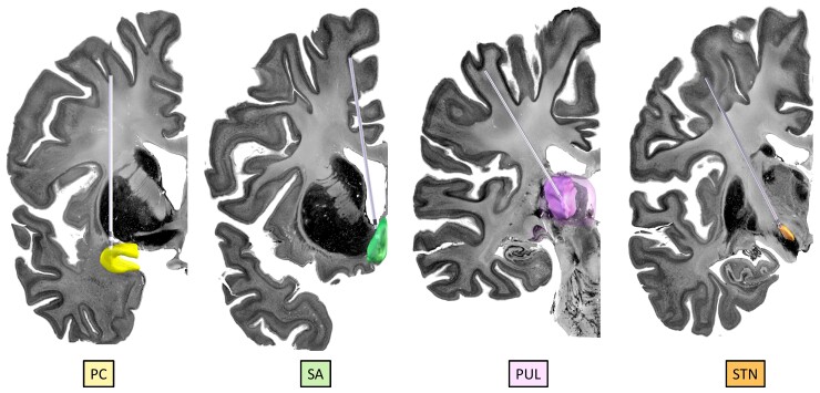
Figure 4.
Demonstration of the anatomical locations of some of the potential propagation points/stimulation targets: The PC (yellow), septal area (SA; green), pulvinar of thalamus (PUL; purple) and STN (orange). The images were created using LeadDBS46,47 with simulated trajectories within the BigBrain backdrop.30 The PC was manually segmented according to the Mai et al. atlas138; the SA was manually segmented; the PUL is a reconstruction from the THOMAS atlas48,49 within LeadDBS and the STN is a reconstruction from the DISTAL atlas139 within LeadDBS.

