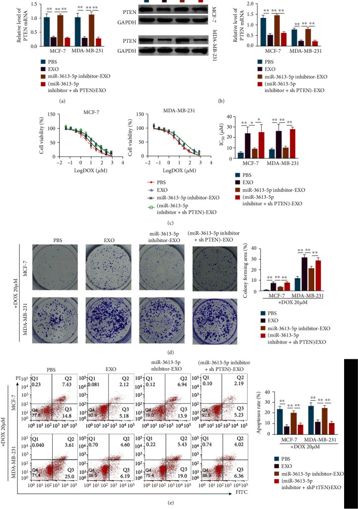
Figure 5.

Exosome-mediated transfer of miR-3613-5p enhances the resistance of breast cancer cells to doxorubicin by targeting PTEN. (a) qRT-PCR was used to assess the relative level of PTEN in MCF-7 and MDA-MB-231 cells after incubation with PBS or exosomes isolated from doxorubicin resistant cells (EXO) and with the treatment of miR-3613-5p inhibitor (miR-3613-5p inhibitor-EXO) and knockdown of PTEN ((miR-3613-5p inhibitor+shPTEN)-EXO). ∗∗p < 0.01. Data are mean ± S.D. of 3 independent experiments. (b) Western blotting was used to detect the protein expression of PTEN in MCF-7 and MDA-MB-231 cells after treatments of PBS or EXO or miR-3613-5p inhibitor-EXO or (miR-3613-5p inhibitor+shPTEN)-EXO. ∗∗p < 0.01. Data are mean ± S.D. of 3 independent experiments. (c) CCK8 was used to assess cell viability of MCF-7 and MDA-MB-231 cells after the cells treated with PBS or EXO or miR-3613-5p inhibitor-EXO or (miR-3613-5p inhibitor+shPTEN)-EXO. (Left and middle) Curve of cell viability after indicated treatments in MCF-7 and MDA-MB-231 cells. (Right) IC50 values of doxorubicin (DOX) after indicated treatments in MCF-7 and MDA-MB-231 cells. ∗p < 0.05, ∗∗p < 0.01. Data are mean ± S.D. of 3 independent experiments. (d) Crystal violet staining to detect colony formation of MCF-7 and MDA-MB-231 cells after the cells treated with 20 μM doxorubicin (DOX) combined with treatments of PBS or EXO or miR-3613-5p inhibitor-EXO or (miR-3613-5p inhibitor+shPTEN)-EXO. ∗∗p < 0.01. Data are mean ± S.D. of 3 independent experiments. (e) Flow cytometry was used to detect the cell apoptosis rate of MCF-7 and MDA-MB-231 cells after the cells treated with PBS or EXO or miR-3613-5p inhibitor-EXO or (miR-3613-5p inhibitor+shPTEN)-EXO. ∗∗p < 0.01. Data are mean ± S.D. of 3 independent experiments.
