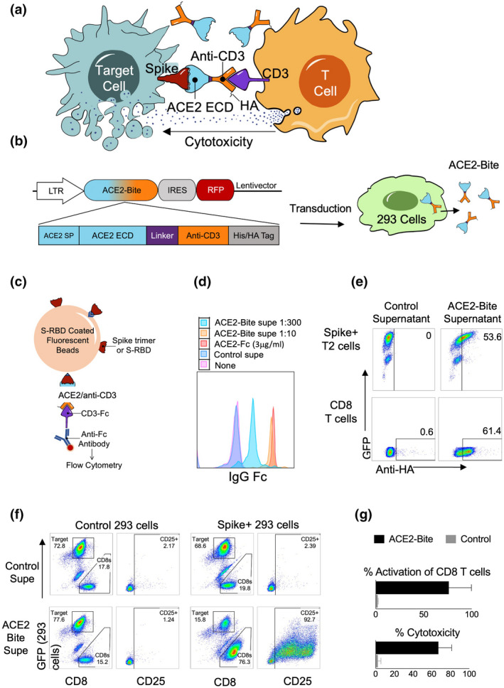
Figure 3.

Functional ACE2/anti‐CD3 bispecific T cell engagers against SARS‐CoV‐2. (a). Mechanism of action of ACE2‐Bite. The extracellular domain (ECD) of ACE2 (blue) in ACE2‐Bite binds to Spike protein (red) expressed on the surface of SARS‐CoV‐2‐infected cells, and the anti‐CD3 fragment (orange) binds to CD3 molecule (purple) on T cells linking both cell types and inducing the activation of T cells, which subsequently results in apoptosis of infected target cells. ACE2‐Bite recombinant protein also contains a hemagglutinin (HA) tag at the C terminal. (b) ACE2‐Bite construct and protein production in 293 cells. A constitutive LTR promoter drives the expression of ACE2‐Bite and RFP genes separated by an Internal Ribosomal Entry Site (IRES). ACE2‐Bite cassette consists of ACE2 signal peptide (SP), ACE2 extracellular domain, a linker peptide, an anti‐CD3 antibody single‐chain variable fragment, a His‐Tag and a Hemagglutinin (HA) Tag. Lentiviruses expressing ACE2‐Bite were used to transduce suspension 293 cells that produce and secrete ACE2‐Bite protein in their culture supernatant. (c) Bead‐based ACE2‐Bite capture assay. Fluorescent beads coated with Spike protein trimer or Spike‐Receptor binding domain (S‐RBD) were used to capture ACE2‐Bite molecules, which were detected via a recombinant CD3‐Fc fusion protein and an anti‐Fc antibody then subsequently analysed by flow cytometry. ACE2‐Fc molecules were also detected with Spike trimer or S‐RBD coated beads and anti‐Fc antibody. (d) Detection of ACE2‐Bite concentrations. 1:10 and 1:300 dilutions were shown in orange and turquoise, respectively, and ACE2‐Fc (3 μg mL−1) (red) using bead‐based ACE2‐Bite capture assay. Wild‐type 293 cell supernatant (Control supe, Blue) and staining buffer (None, Pink) were used as negative controls. (e) Binding of ACE2‐Bite to Spike‐GFP‐expressing T2 cell line and primary human T cells. HA staining of Spike‐GFP‐expressing T2 cells (top panel) and CD8 T cells (bottom panel) when combined with ACE2‐Bite (right plot) or control (non‐transduced 293) (left plot) supernatants. (f) CD25 and GFP expressions show activation and cytotoxicity of resting CD8 T cells against Spike/GFP‐expressing or control (transduced with GFP‐expressing empty vector) 293 cells in the presence of ACE2‐Bite or control supernatant. (g) The bar graph demonstrating the results of the cytotoxicity assay of the experiment is represented in f. The experiments were replicated three times with similar results.
