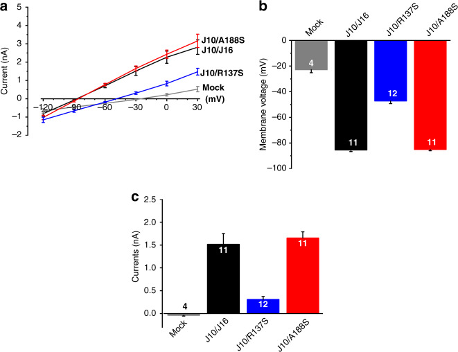
Fig. 2. Electrophysiological characterization of KCNJ16 variants.
a Current–voltage curves of human embryonic kidney cells (HEK293T) either non-transfected (mock; gray line) or co-transfected with KCNJ10 (J10) and KCNJ16WT (J16, black line) or mutant KCNJ16 (R137S, blue line; A188S, red line). The variant R137S seemed non-functional, whereas A188S was fully functional. Data are presented as mean values ± SEM. b Resting membrane potential in HEK293T cells: KCNJ10/KCNJ16 (J10/J16, black bar) and KCNJ10/KCNJ16A188S (J10/A188S, red bars) transfected cells showed hyperpolarized membrane potentials close to the equilibrium potential for K+ (approximately −90 mV). Membrane potential of KCNJ10/KCNJ16R137S (blue bar) transfected HEK293T cells were depolarized, like the non-transfected cells (gray bar). Data are presented as mean values ± SEM, numbers indicate the numbers of experiments. c Whole-cell current of HEK293T clamped at −30 mV: KCNJ10/KCNJ16 (J10/J16, black bar) and KCNJ10/KCNJ16A188S (J10/A188S, red bar) transfected cells showed nearly 4-times higher whole-cell current than KCNJ10/KCNJ16R137S (J10/R137S, blue bar). Data are presented as mean values ± SEM, numbers indicate the numbers of experiments. (For interpretation of the references to color in this figure, the reader is referred to the online version of this article.)

