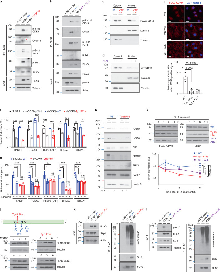
Fig. 5. The p-Tyr19-CDK9 is important for its kinase activity and protein stability.
a, Expression of the indicated proteins in SKOV3-stable cells expressing Tyr19Phe and WT CDK9 examined by WB after IP with FLAG antibody. Data represent two repeats with similar results. b, Expression of the indicated proteins in SKOV3-stable cells expressing WT CDK9 with or without ALK inhibitor treatment examined by WB after IP with FLAG antibody. Data represent two repeats with similar results. c,d, Subcellular localization of FLAG-tagged CDK9 in cells expressing Tyr19Phe (c) and WT CDK9 treated with or without ALK inhibitor (d). Data represent two repeats with similar results. e, Representative images of FLAG–CDK9 with DAPI staining (upper panel) and quantification of cells with nuclear CDK9-positive signal (lower panel) in cells expressing Tyr19Phe CDK9 and WT CDK9 treated with or without ALK inhibitors. Error bars represent mean ± s.e.m. of n = 3 independent experiments. Scale bar, 20 μM. f, Quantitative PCR analysis of gene expression in CDK9-knockdown SKOV3 cells rescued with WT or Tyr19Phe CDK9. Error bars represent mean ± s.d. of n = 3 independent experiments. g, Quantitative PCR analysis of gene expression in CDK9-knockdown SKOV3 cells rescued with WT or Tyr19Phe CDK9 treated with or without ALK inhibitors. Error bars represent mean ± s.d. of n = 3 independent experiments. h, WB of HR factors and PARP1 level in cells expressing WT or Tyr19Phe CDK9 after treatment with or without 0.5 μM ALK inhibitor (lorlatinib) for 24 h. Data represent two repeats with similar results. i, WB of FLAG-tagged CDK9 in SKOV3-stable cells expressing Tyr19Phe CDK9 and WT CDK9 treated with or without ALK inhibitors. Cells were treated with 50 μΜ CHX for the indicated time (upper panel). Quantification of band intensity is shown in the lower panel. Error bars represent mean ± s.e.m. of n = 3 independent experiments. j, WB of FLAG-tagged CDK9 in SKOV3-stable cells expressing Tyr19Phe or WT CDK9. Cells treated with 10 μΜ proteasome inhibitors (MG132 or PS-341) for the indicated time. Data represent two repeats with similar results. k,l, Expression of ubiquitination and Skp2 examined by WB after IP with FLAG antibody. k, SKOV3 cells stably expressing WT or Tyr19Phe CDK9 treated with MG132 (10 μΜ). l, SKOV3 cells stably expressing WT CDK9 treated with or without ALK inhibitor (lorlatinib, 0.5 μM) and MG132 (10 μΜ). Data represent two repeats with similar results. Statistical analysis was carried out using the two-tailed, unpaired Student’s t-test (e) or two-way ANOVA (f, g and i). NS, not significant. *P < 0.05, **P < 0.01, ***P < 0.001.

