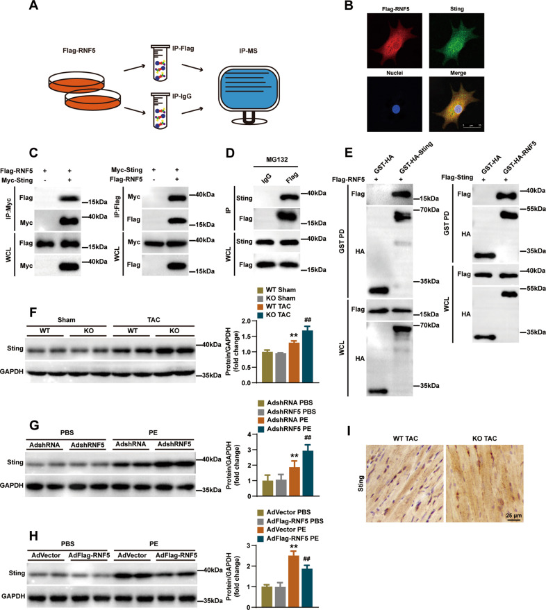
Fig. 6. The interaction between STING and RNF5 was confirmed.
A The flow chart shows the Co-IP and subsequent IP-MS of RNF5, screening for candidate proteins that interact with RNF5. B NRCMs was infected with AdFlag-RNF5 after MG132 was added 6 h before harvest, and immunofluorescence showed that Flag-RNF5 (red) co-located with STING (green). C After HEK-293T cells were transfected with Flag-RNF5 and Myc-STING plasmids, Co-IP detection suggested that RNF5 and STING interacted. D After NRCMs were infected with AdFlag-RNF5 and IgG antibody was used as a negative control, MG132 was added 6 h before harvest. Co-IP demonstrated the interaction between exogenous RNF5 and endogenous STING. E Glutathione S-transferase precipitation assay showed that RNF5 is directly bound to STING. F WB analysis of STING protein levels in WT and RNF5 KO mice 4 weeks after TAC or sham surgery. G WB analysis of STING in NRCMs infected with AdshRNA or AdshRNF5 after 24 h treatment with PBS or PE. H WB analysis of STING in NRCMs infected with AdVector or AdFlag-RNF5 after 24 h treatment with PBS or PE. I Immunohistochemical showed the expression of STING in the indicated groups. Scale bars: 50 μm. For (F–H) **P < 0.01 versus WT Sham or AdshRNA PBS or AdVector PBS, ##P < 0.01 versus WT TAC or AdshRNA PE or AdVector PE. Data were presented as the mean ± SD. Statistical analysis was carried out by one-way ANOVA.

