Fig. 1.
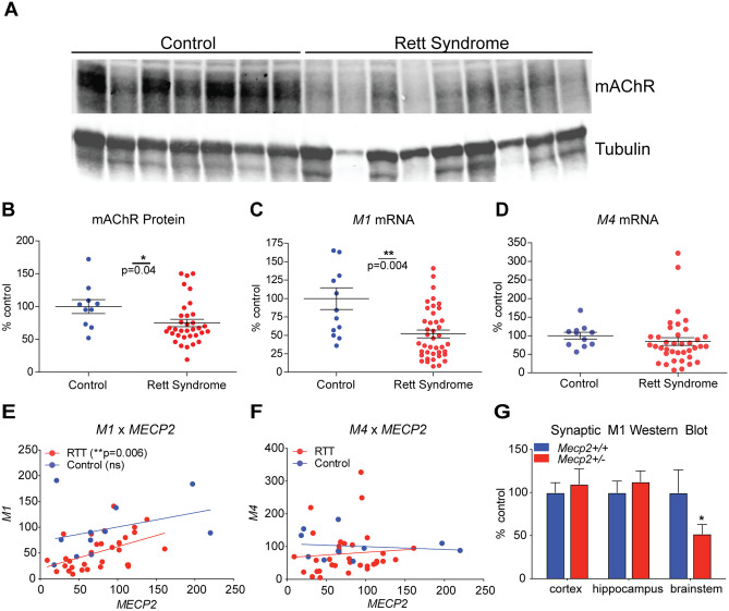
M1 expression is decreased in temporal cortex samples from 40 Rett syndrome (RTT) autopsy samples. A–B Western blot. mAChR (M1–M5) protein levels are significantly decreased in the brain samples (BA38) from RTT autopsies when compared relative to age, sex, and postmortem interval matched controls (N = 12). Student’s t test. C–D qRT-PCR. mRNA expression of the mAChR subtype M1 receptor is significantly decreased in human RTT samples, while M4 levels are comparable to controls in the temporal cortex. Student’s t test. E–F Linear regression analysis. A correlative analysis shows a significant linear relationship between M1 and MeCP2 transcript levels, which is not observed with M4. Linear regression. G Western blot. Relative to Mecp2+/+ controls, synaptosome preparations from 20-week-old Mecp2+/- mice show a significant reduction in mAChR levels in the brainstem. Two-way ANOVA with Tukey post hoc analysis. N = 5/treatment/genotype. *p < 0.05, **p < 0.01

