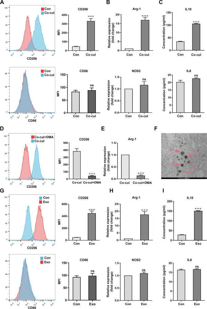
Fig. 4. SCLC cells promote the M2 polarization of BMDMs by secreting exosomes.
A Flow cytometry of the expression levels of the M2 polarization marker CD206 and M1 polarization marker CD86 in BMDMs without or with RT SCLC cell co-culture (n = 3). B qRT-PCR analyses of the expression levels of the M2 polarization marker Arg-1 and M1 polarization marker NOS2 in BMDMs (n = 3). C ELISA results of IL10 and IL6 in supernatant of BMDMs (n = 3). D, E Analysis of BMDMs, co-cultured with SCLCs post DMA treatment. D Flow cytometry of the expression levels of CD206 in BMDMs in a co-culture system with or without DMA treatment (n = 3). E qRT-PCR analyses of the expression levels of Arg-1 in BMDMs (n = 3). F A TEM image of exosomes isolated from RT SCLC cell culture medium. Scale bar, 200 nm. G–I Analysis of BMDMs cultured with SCLC-derived exosomes. G Flow cytometry of the expression levels of CD206 and CD86 in BMDMs treated without or with SCLC-derived exosomes (n = 3). (H) qRT-PCR analyses of the expression levels of Arg-1 and NOS2 in BMDMs (n = 3). I ELISA results of IL10 and IL6 in supernatant of BMDMs (n = 3). Con, untreated BMDMs. Co-cul, BMDMs co-cultured with RT SCLC cells. Exo, BMDMs co-cultured with SCLC-derived exosomes. Bar graphs show the mean ± SEM; ***P < 0.001. SCLC small cell lung cancer, BMDMs bone marrow-derived macrophages, DMA dimethyl amiloride, TEM transmission electron microscopy, SEM standard error of mean.

