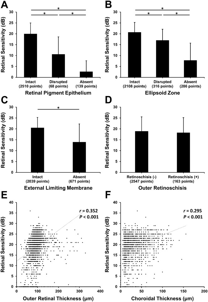
Figure 2.
(A–D) Retinal sensitivity according to the integrity of hyperreflective lines of RPE (A), EZ (B), and ELM (C) and the presence of outer retinoschisis (D) on OCT. Asterisks indicate statistically significant differences. (E, F) Scatterograms showing the correlation of retinal sensitivity with ORT (E) and CT (F).

