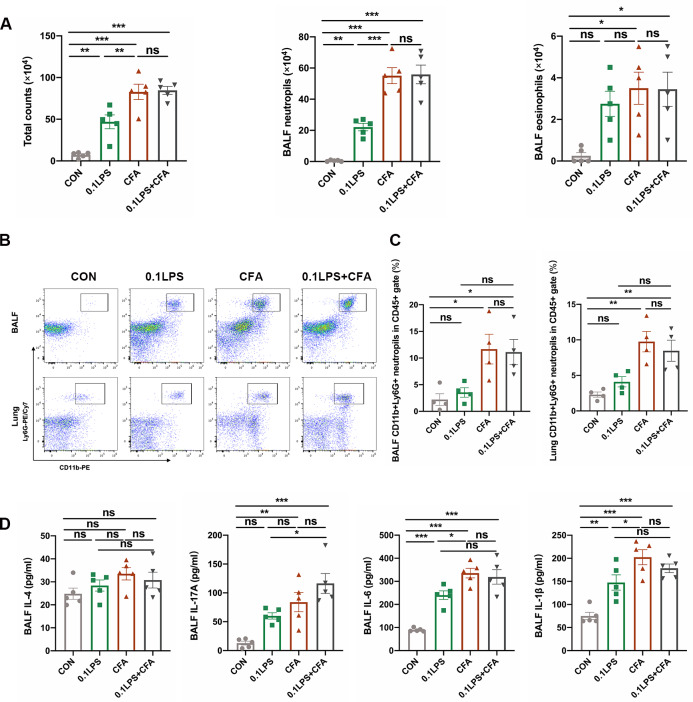
Fig. 5.
Significant neutrophilic inflammation and secretion of proinflammatory factors were observed in CFA combined with LPS induced mouse model. (A) Quantification of total cell counts and differential cell counts (eosinophils and neutrophils) in BALF at 48 h after the last challenge. (B) The percentage of CD11b(+)Ly6G(+) neutrophils in CD45(+) leucocytes in BALF and lung tissue was determined by flow cytometry at 48 h after the last challenge. The representative images of each group are shown. (C) Statistical analysis of the above data shown by flow cytometry. (D) Cytokine (IL-4, IL-6 and IL-1β) concentrations in BALF were quantified by enzyme-linked immunosorbent assay (ELISA). Data were shown as mean ± SEM; n = 5 in (A) and (D); n = 4 mice in (B) and (C). Significance between groups was calculated using one-way ANOVA with Tukey’s post hoc method. *p < 0.05, **p < 0.01 and ***p < 0.001

