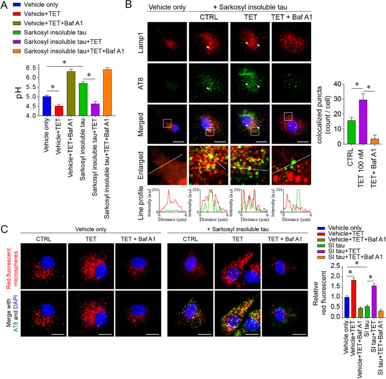
Fig. 7.
Tetrandrine restores the microglial phagocytosis impairment induced by pathological tau. A Lysosomal pH measurements in primary microglia 24 h after incubation with Sarkosyl-insoluble fractions from tau-P301S mouse brain homogenates with or without tetrandrine treatment. The Sarkosyl vehicle was used as the control. BafA1 was used to induce lysosomal alkalinization. Data are summarized as the mean ± SEM from 3 independent experiments. *Indicates p < 0.05. B Representative micrographs showing the localization of phagocytosed tau in microglia. Lamp1 and AT-8 were employed to label lysosomes and phosphor-tau, respectively. The enlarged images show the magnified region in the white boxes from the merged images. White arrowheads indicate the colocalization of tau and Lamp1, and their degree of colocalization was analysed with ImageJ with the colocalization plugin. The red and green traces in the line profile show the intensity of red and green colour in arbitrary units (a.u.) along the white line indicated in the enlarged images. Data are summarized as the mean ± SEM from 15 images analysed in each group. *Indicates p < 0.05. C Representative micrographs showing the phagocytosis assay of red fluorescent microspheres under the influence of tetrandrine (TET) and bafilomycin A1 (BafA1) in primary microglia 24 h after incubation with Sarkosyl-insoluble (SI) fractions of P301S tau mouse brain homogenates or Sarkosyl vehicle only. Data are summarized as the mean ± SEM from 15 images analysed in each group. *Indicates p < 0.05

