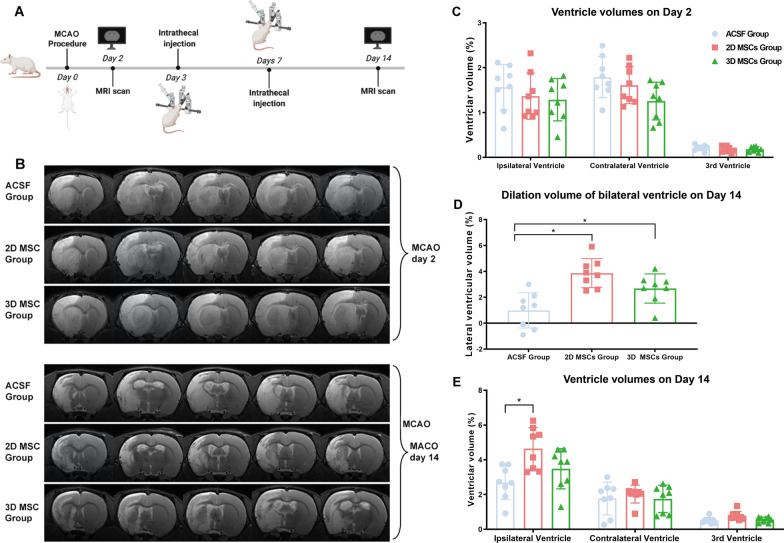
Fig. 2.
Ventricular enlargement in MCAO rats after intrathecal injection of MSCs. A Experimental scheme showing the times of MCAO surgery, MRI analysis and intrathecal injection of MSCs (1 × 10⁶ per dose) in rats. B MRI images showing the regions of cerebral infarcts and changes in lateral ventricular sizes before MSC transplantation (Day 2 post-MCAO) and after MSC transplantation (Day 14 post-MCAO; n = 8; representative images of one rat from each group are shown). C–E Measurement of ventricular sizes by MRI, indicated by ventricular volume as a percentage of bilateral hemisphere volume, showed no significant differences among the three groups on Day 2 before MSC transplantation (C). There was a significant increase in the size of the ipsilateral and the third ventricle in 2D MSC-treated rats but not in 3D MSC-treated rats, compared to ACSF-treated controls at 14 days (D; P < 0.05; n = 8). And the dilation volume of lateral ventricle in rats receiving MSCs was significantly higher than that in rats receiving ACSF (E; *P < 0.05; n = 8)

