
FIGURE 2.
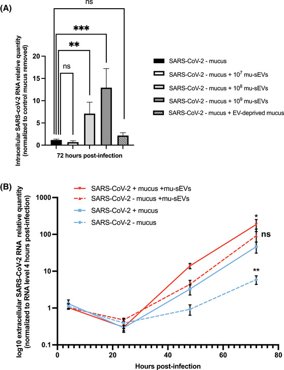
Effect of mu‐sEVs and mucus‐containing sEVs on HNEC infection by SARS‐CoV‐2. (A) Effect of preincubation of viral particles with different concentrations (107–109) of mu‐sEVs or with EV‐deprived mucus on SARS‐CoV‐2 infection (10 μl of viral inoculum, ∼2.04 × 10⁵ TCID50/ml) at the apical pole of HNECs after removal of recipient cell mucus. Intracellular SARS‐CoV‐2 RNA was extracted 72 h post‐infection and quantified by RT‐qPCR. Results were normalized to 18S rRNA, then to mucus‐free control and viral particle preincubation with PBS alone (black bar). They are expressed as mean ± SEM of two independent experiments. **p < 0.01, ***p < 0.001. (B) Dynamics of SARS‐CoV‐2 RNA production at the apical pole of HNECs, as assessed by RT‐qPCR. Results were normalized to the viral RNA level 4 h post‐infection for each condition and expressed as log10 mean ± SEM of two independent experiments. *p < 0.05, **p < 0.01 (Mann‐Whitney U‐test versus extracellular SARS‐CoV‐2 RNA levels in the presence of recipient cell mucus 72 h post‐infection (blue line))