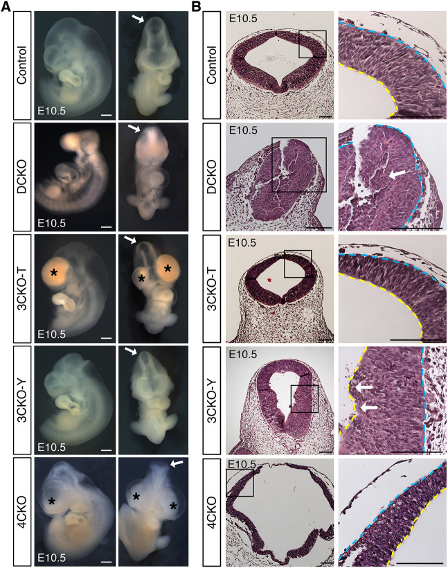
Fig. 7.
Hippo signaling maintains neuroepithelial cell behavior in the dorsal neural tube. (A) Bright-field images of E10.5 rescue embryos. 3CKO-T and 3CKO-Y embryos showed comparable midbrain morphology to that of control (arrows). 3CKO-T and 4CKO embryos had forebrain and pharyngeal arch defects (asterisks). Scale bars: 500 μm. (B) H&E staining of coronal sections. Neuroepithelium integrity is recovered in 3CKO NTs. Some apical extrusion into the ventricle is detected in 3CKO-Y NTs (arrows), like the infiltration sites in DCKO NTs. Boxed areas are shown at a higher magnification (right). Scale bars: 100 μm. Yellow lines indicate apical edge; blue lines indicate basal edge.

