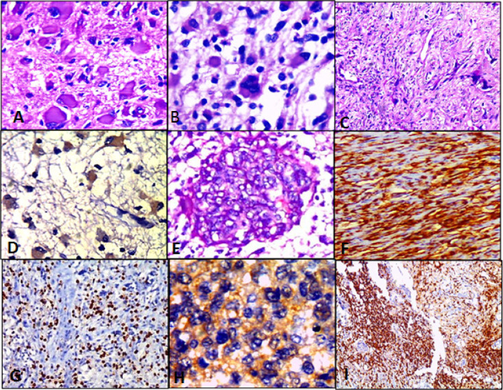
Figure 2.
Different Cases of High Grade Gliomas; Figures (A, B, E &C); Anaplastic astrocytoma with neoplastic gemistiocytes, WHO grade III; Glioblastoma, WHO grade IV; Micovascular proliferation & Gliosarcoma, WHO grade IV respectively (H&E; (A)X400, (B)X400, (E)X 200 & (C)X100 original magnification). Figures (D, H & F); Anaplastic astrocytoma shows LC3B brown cytoplasmic staining in about 70% of tumor cells, Score 9+; Glioblastoma shows LC3B tan cytoplasmic staining in about 80% of tumor cells, Score 8+ and Gliosarcoma shows LC3B brown cytoplasmic staining in about 90% of tumor cells, Score 12+ respectively (IHC; (D)X400, (H)X400 & (F)X200 original magnification). Figures (G & I); High grade gliomas show dense CD3 positive lymphocytes scattered between tumor cells (IHC; (G)X200 & (I)X100 original magnification)

