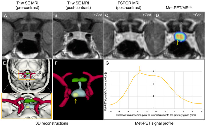
Fig. 1.
MRI and Met-PET findings with 3D reconstruction of the sella and parasellar regions in a patient with ACTH-dependent Cushing’s syndrome. A–C Pre- and post-contrast coronal T1w SE MRI (A, B) and FSPGR (volumetric) MRI (C) demonstrate equivocal appearances, with subtle deviation of the infundibulum to the left, but minor downward sloping of the floor of the sella on the left side. No discrete microadenoma is readily visualized. D Met-PET/MRCR reveals both central (white arrow) and right-sided (yellow arrow) radiotracer uptake in the gland. E, F 3D reconstructed images, combining PET, CT and FSPGR MRI datasets, allows appreciation of the location of the tumor (yellow) with respect to the normal gland (turquoise) and other adjacent structures including the intracavernous carotid arteries (red) and optic chiasm (green). G Profiling of 11C-methionine uptake across the sella reveals two peaks consistent with uptake by normal gland and a corticotroph microadenoma. At transsphenoidal surgery, a microadenoma was resected from the right side of the gland and confirmed histologically to be a corticotroph adenoma. Postoperatively the patient achieved complete clinical and biochemical remission and remains eupituitary. Key: CT computed tomography; FSPGR fast spoiled gradient recalled echo; Gad gadolinium; Met-PET/MRCR 11C-methionine PET-CT coregistered with volumetric (FSPGR) MRI; MRI magnetic resonance imaging; PET positron emission tomography; SE spin echo; T1w T1-weighted

