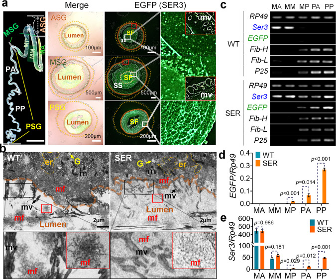
Fig. 4. Synthesis and secretion of silk proteins in SGs of mutant 5th instar larvae.
a Frozen section of the SG cross-section. The state of the SER3 protein secreted into the lumen of the SG, was detected by EGFP-fusion expression. b Transmission electron micrograph of the PSG. Fig. 4a and b: SF, silk fibroin layer; SS, silk sericin layer; er, endoplasmic reticulum; G, Golgi apparatus; m, mitochondrion; mf, fibroin mass; mv, microvilli. c Semi-quantitative PCR and d, e qRT-PCR to detect the mRNA levels of EGFP, SER3, and silk fibroin Fib-H, Fib-L, and P25 genes in cells in different parts of the SG. MA, MM, and MP show the anterior, middle, and posterior parts of the MSG, respectively. PA and PP show the anterior and posterior parts of the PSG, respectively. For d and e, Holm–Sidak t-test analysis was used and the p value obtained was the adjusted p value. Data were presented as mean ± SEM. n = 3 samples. Each tissue sample was collected from three female individuals, and each sample was measured three times. Image data are representative of three independent experiments unless otherwise stated.

