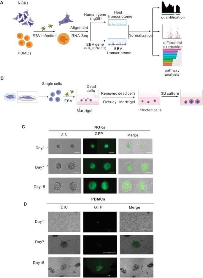
Fig. 1. Establishment of EBV-infected NOK-derived 3D culture system and PBMC.
Generation and confirmation of EBV infection. To establish 3D NOKs cultures, Matrigel was poured onto 12-well culture plates to a depth of approximately 0.2 mm, followed by polymerization for 30 min at 37 °C. The mixture of BAC GFP-EBV and single-cell suspension of NOKs were placed on the surface of the Matrigel matrix and incubated at 37 °C overnight. The next day dead cells were removed by aspiration, and a Matrigel layer was overlaid to cover the cells attached to Matrigel at the bottom. After the Matrigel solidified, the culture medium was added and changed every 2 days. After 2 days the NOKs cells invade the matrix to form multicellular spheroids. PBMCs were infected with BAC GFP-EBV, then we check the GFP after 7 days and 15 days. A Schematic illustration of study design. B The flowchart of key procedures to establish BAC GFP-EBV infected NOKs-derived 3D culture system. C Representative image of BAC-GFP-EBV infection-mediated GFP expression in NOKs 3D culture at different days (amplification: 400×). D Representative image of BAC-GFP-EBV infection-mediated GFP expression in PBMCs culture at different days (amplification: 200×).

