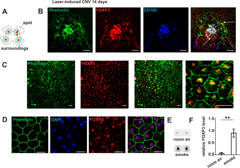
Fig. 4.
FoxP3 expression in choroidal neovascularization and after long-term smoke exposure in mice. Laser-induced choroidal neovascularization (CNV) in a mouse eye, flatmount preparation of the RPE prepared 14 days after laser burn. A Scheme of flatmounted RPE/choroid illustrating the arrangement of laser burns, peri-lesion area (spot) and periphery (surroundings). B Immunofluorescence staining (as indicated) of a mouse RPE flatmount with a laser scar showing RPE cells with distorted borders (phalloidin green), FoxP3 (red) positive cells marked with white arrows and blood vessels (CD102 blue). Scale bar represents 50 µm. C RPE structure and FoxP3 expression in the peri-lesion area (three left panels) and at higher magnification (right panel); FoxP3 in red and phalloidin in green. Scale bar represents 20 µm. D Localization of FoxP3 in the nucleus in the laser CNV model; phalloidin (green), DAPI (blue) and FoxP3 (red) verify the localization of FoxP3 in the nucleus. Scale bar represents 20 µm. E, F FoxP3 levels under the influence of cigarette smoke in mice. Mice have been exposed to cigarette smoke for 6 months according to Woodell et al. [88]. E Dot blots for FoxP3 from extracts of RPE/choroid. F Quantification of FoxP3 levels. Dot blots of RPE/choroid extracts were probing with anti-mouse FoxP3 antibody, using GAPDH for normalization (**p < 0.01; N = 3 per condition)

