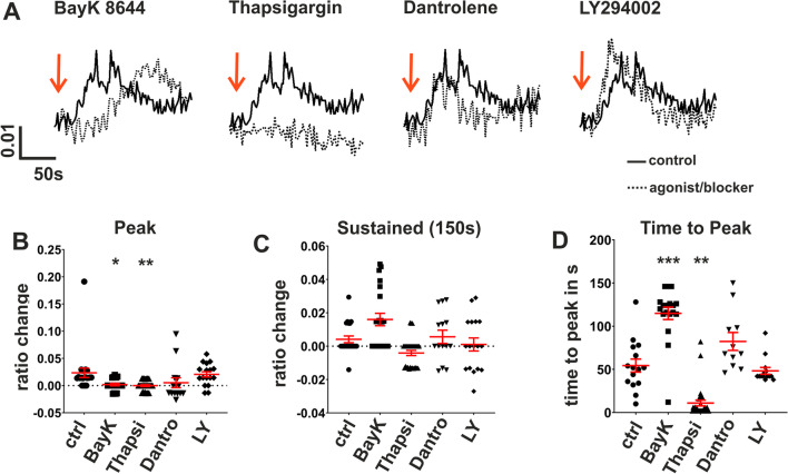
Fig. 7.
Analysis of IL-1β-evoked Ca2+-transients in non-confluent ARPE-19 cells. Intracellular free Ca2+ was measured in non-confluent ARPE-19 cells using the Ca2+-sensitive fluorescence dye Fura-2; changes in intracellular Ca2+ were plotted as changes in the fluorescence ratio of the excitation wavelengths. A Representative raw data from single-cell experiments (solid line represents IL-1β (100 ng/ml) alone; dotted lines IL-1β after 5 min preincubation with the following inhibitors of Ca2+-signaling: 10 µM BayK 8644 (blocker of L-type Ca2+ channels), 1 µM thapsigargin (blocker of sarcoplasmic Ca2+-ATPase to empty intracellular Ca2+-stores); 1 µM dantrolene (blocker of ryanodine receptors); 50 µM Ly294002 (blocker of PI3-kinase). B Comparison of IL-1β-evoked Ca2+ sustained elevation. C Comparison of IL-1β-evoked Ca2+-peaks. D Comparison of time-to-peak of IL-1β-evoked Ca2+ transients. (BayK = BayK8644; Thapsi = thapsigargin; Dantro = dantrolene; LY = Ly294002). Data are presented as mean values ± SEM. Statistical significance was calculated using Mann–Whitney U test

