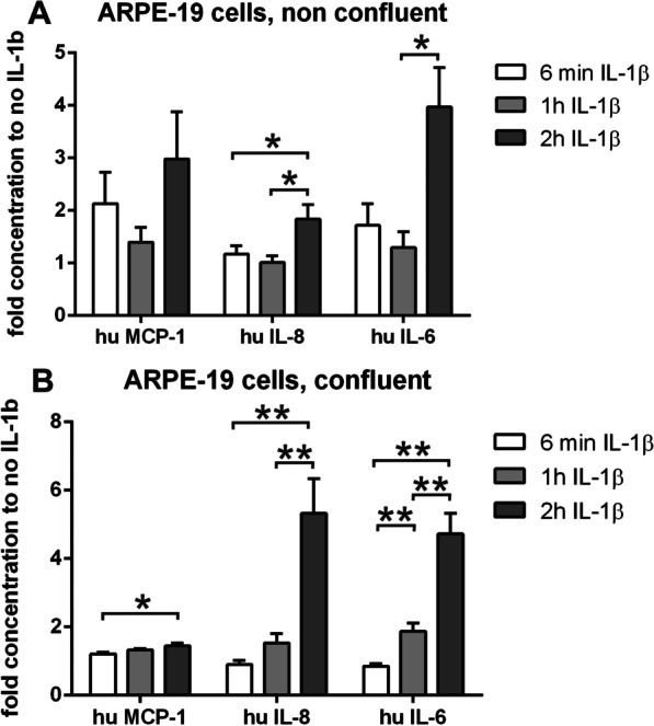
Fig. 9.

Secretion of cytokines and chemokines by ARPE-19 cells after stimulation with IL-1β. Secretion of MCP-1, IL-8 and IL-6 (pg/ml) by ARPE-19 cells stimulated with IL-1β for 6 min, 1 and 2 h. A Non-confluent cells. B Confluent cells (IL-6 = interleukin-6; IL-8 = interleukin-8; MCP-1 = monocyte chemoattractant protein-1/CCL2). Triplicate cultures of ARPE-19 were treated as described; culture supernatants were collected at the end points of IL-1β-treatment. Supernatants were pooled and tested for cytokine and chemokine secretion as duplicates in a multiplex bead assay. The final values of duplicate supernatants are calculated by the standard curves of the assay from the median fluorescence intensity of at least 50 beads measured for each analyte and sample and normalized to the cell number of confluent cultures (cytokine concentration of non-confluent cultures × 1.5). The data show the x-fold concentrations (means + SEM) of cytokines compared to the unstimulated cultures (*p < 0.05; **p < 0.01; N = 4–6)
