Fig. 8.
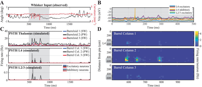
Network response to in vivo-like stimulation. (A) Input to the network: whisker angle (black) and curvature (red) from a freely whisking rat in a pole localization task (data from (Peron et al., 2015), made available as 'ssc-2' on CRCNS.org). (B) Example voltage trace responses of 6 randomly chosen model neurons. (C) Peri-Stimulus Time Histograms (PSTHs) of the model-thalamus (top), L4 (middle) and L2/3 (bottom). The thalamus consists of 3 barreloids, each containing 200 'filter-and-fire' neurons that respond to whisker angle, curvature or a combination of both. The central barreloid (black, 2) receives a stronger input, as this is the 'stimulated' barrel for the only spared whisker. Spike trains of the thalamus are sent to the cortical network model of L4 (middle), which sends its spike trains to L2/3 (bottom). These similarly consist of 3 barrels, of which the central (black, 2) barrel belongs to the spared whisker. (D) Average membrane potential of the excitatory (left) and inhibitory (right) model neurons as a function of cortical depth. L4 (barrel cortex) is denoted with a grey shaded shape. (E) Average firing rates of the model neurons as a function of cortical depth

