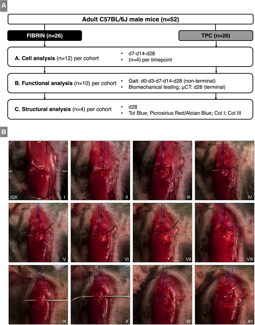
Figure 1: Study design and intraoperative imaging of the surgical procedure.
(A) Experimental study design and treatment groups. (B) Surgical procedure for supraspinatus tendon detachment, fibrin gel delivery, and repair. (I) Anatomic overview after skin incision. (II) Horizontal incision of deltoid. (III) Additional exposure was obtained for imaging purposes to visualize the subscapularis, supraspinatus and infraspinatus tendons. (IV-VII) The supraspinatus tendon is secured with a modified figure 8 stitch to obtain equal pulling tension on the tendon. (VIII) Sharp detachment of the tendon from the greater tuberosity. (IX) Posterior to anterior bone tunnel. (X) Suture is passed via the 30G needle from anterior to posterior. (XI) Suture is tied and tensioned to closely approximate the native insertion site. (XII) Fibrin gel bead (white arrow) is added to the enthesis region. BioM: Biomechanical testing; μCT: Micro-CT imaging; T: Trapezius muscle; ACR: Acromion; D: Deltoid muscle; SSc: Subscapularis tendon; SS: Supraspinatus tendon; IS: Infraspinatus tendon; GT: Greater tuberosity. Images were taken at 10x magnification.

