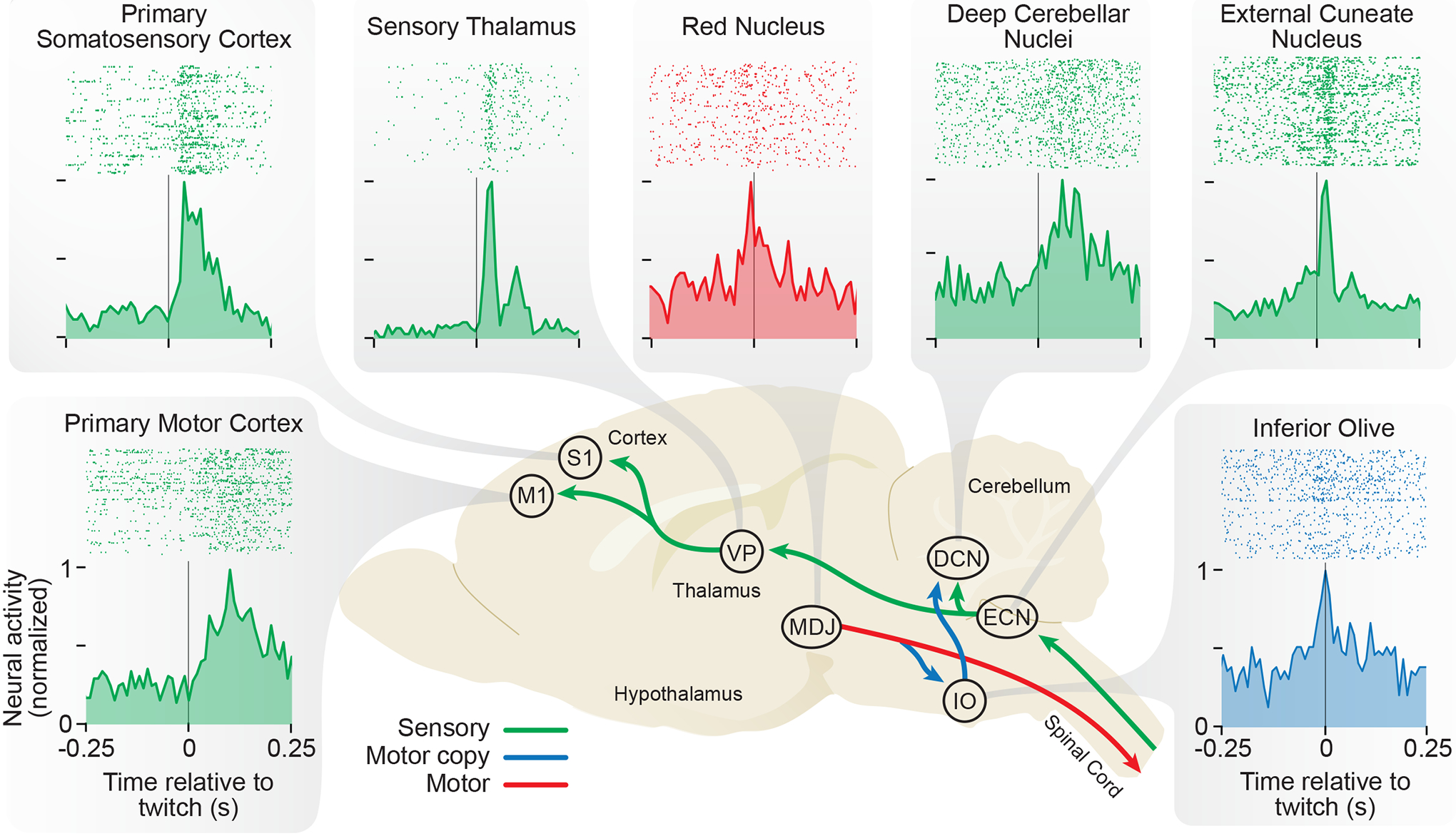
Figure 4.

Diversity of Twitch-Related Neural Activity In the Infant Rat Brain
Representative perievent histograms depicting neural activity recorded from P8 rats in relation to forelimb twitches. The background image depicts a sagittal section of the infant brain. The production of a forelimb twitch begins in the brainstem, including the red nucleus and other motor structures within the mesodiencephalic junction (MDJ). The forelimb movement activates proprioceptors and triggers sensory feedback (reafference) that flows to the external cuneate nucleus (ECN) in the medulla, the ventral posterior nucleus (VP) in the thalamus, and primary somatosensory (S1) and primary motor (M1) cortex. The RN and adjacent motor neurons also convey motor copies (corollary discharge) to the inferior olive (IO) that, in turn, projects to the deep cerebellar nuclei (DCN). For clarity, not all structures known to exhibit twitch-related activity are shown. Arrows and histograms are color-coded based on whether they reflect motor commands (red), motor copies (blue), or sensory feedback (green). See text for references.
