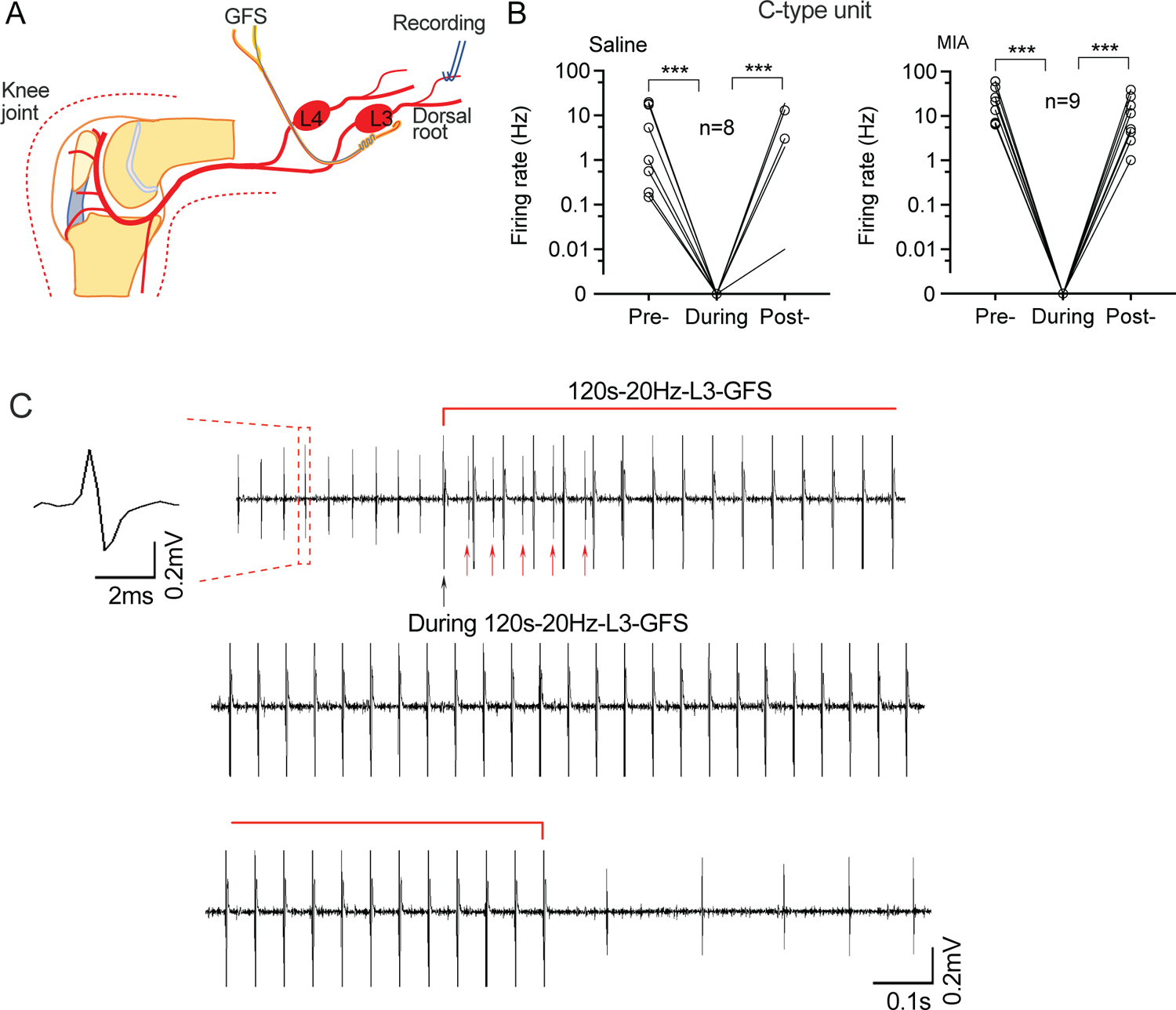
Figure 2.

Effects of 120s-20Hz GFS on SA in L3 dorsal root units. (A) Preparation design. (B) SA recorded from C-type units in the L3 dorsal root is reduced during (the first 10s following) L3 GFS. (C) Representative trace from a C-type unit showing blocked SA soon after the start of GFS (top), during (middle), and after GFS (bottom). Black arrow points to the stimulation artifact of the first stimulation of GFS. Red arrows point to SA during the first several stimulations of 20Hz GFS. Data are from n=4 rats with saline injection and n=4 rats with injection of MIA. *** P<0.001 with nonparametric 1-way repeated measure ANOVA (Friedman test) followed by comparison of rank.
