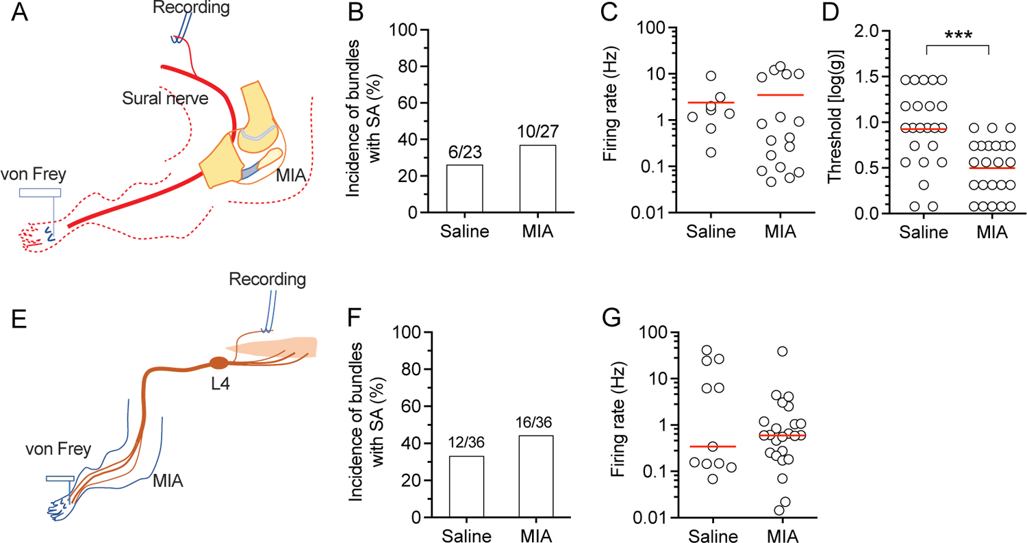
Figure 4.

SA in sural nerve and L4 dorsal root units with the RFs in the plantar skin. (A) Preparation design for recording from the sural nerve. (B-D) Graphs show the frequency of SA in bundles (B), SA firing rate (C), and the threshold of mechanical stimulation of the RF in the hind paw for inducing firing of units in sural nerve teased bundles (D). (E) Preparation design for recording from the L4 dorsal root. Graphs show the frequency of bundles with SA (F) and firing rate (C). Red lines represent means. Data are from n=5 rats with saline injection and n=5 rats with injection of MIA for each type of recordings from sural nerve, and L4. *** P<0.001 by t-test.
