Figure 8.

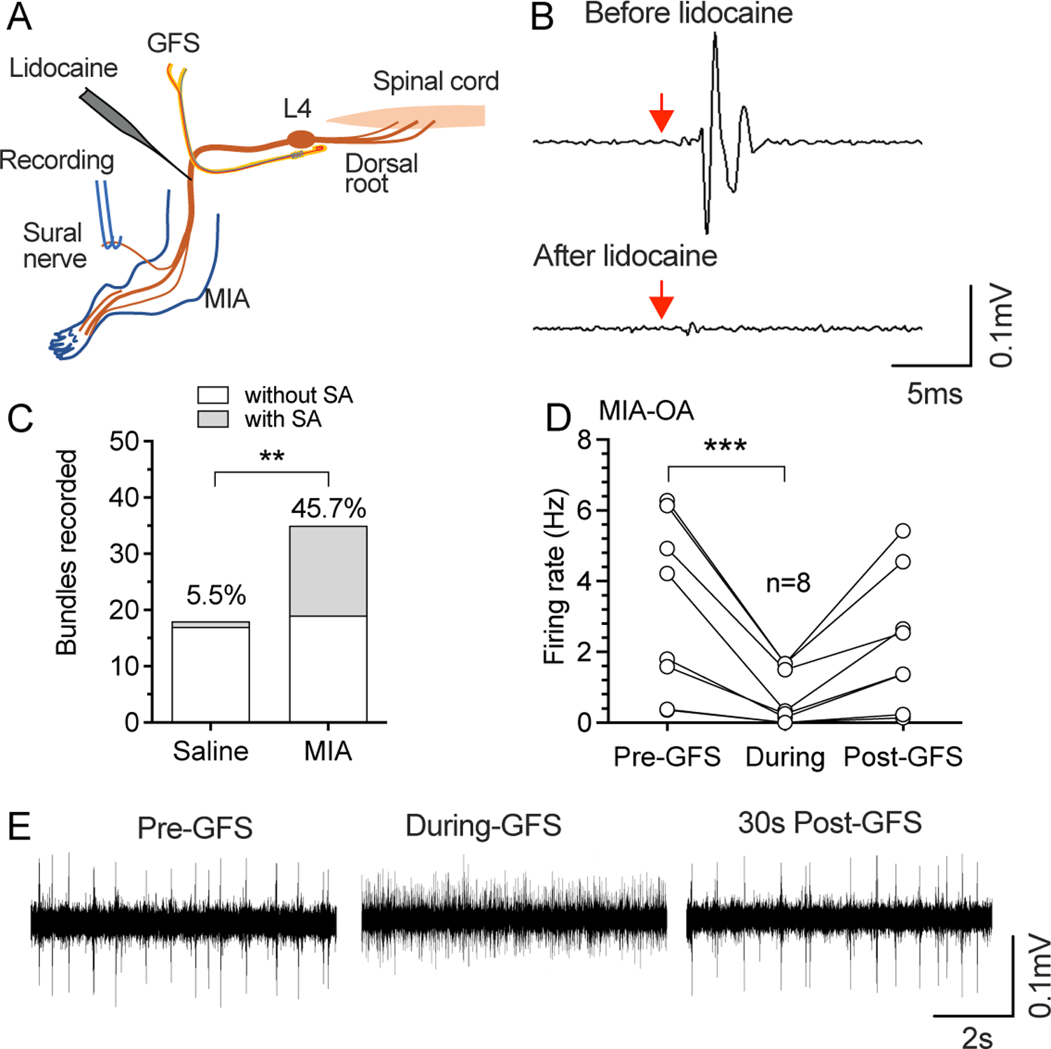
Retrograde SA recorded from the sural nerve. (A) Preparation design for recording efferent activity from the sural nerve. (B) Sample traces show retrograde firing evoked by single stimuli from the GFS electrode before (top) and after (bottom) application of TTX to sciatic nerve, which was used to identify recordable single units, and thereafter measure their SA. Red arrows point to the start of stimulation. (C) MIA-OA increased the frequency of retrograde SA recorded from sural nerve. ** P<0.01 by Chi-square comparisons. (D) 120s-20Hz GFS blocked the incidence of retrograde SA recorded from sural nerve after MIA-OA. ***P<0.001 nonparametric 1-way repeated measure ANOVA (Friedman test) followed by comparison of rank. (E) Sample traces shows effects of GFS on retrograde SA. These recordings were from 5 rats with saline injection and n=5 rats with injection of MIA.
