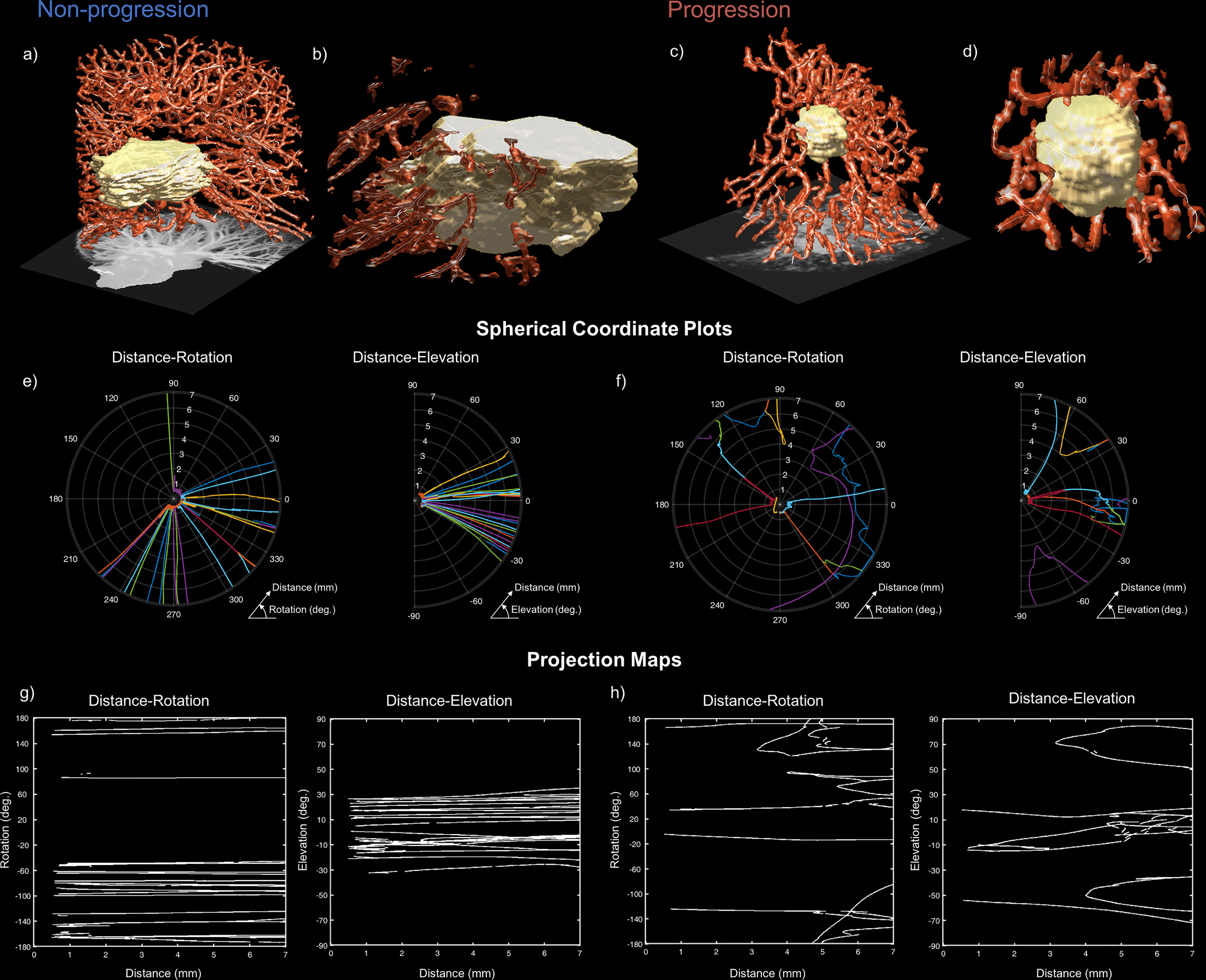
Figure 4.

Organization of vascular network at the tumor interface distinguishes NSCLC tumors that experience durable response (left) from those that progress (right) following platinum-based chemotherapy (NSCLC-PLAT). High vascular density is observed in both non-progressors (a) and progressors (c), but differences in the arrangement of tumor-adjacent (b and d) vessels are detectable through QuanTAV Spatial Organization features. e-h) On projection images depicting rotation around the tumor centroid (left) and elevation above the tumor centroid (right) with respect to distance from the tumor, the standard deviation of vessel orientation was elevated among patients who experienced progression. The position of vessels in a spherical coordinate system relative to the tumor, depicted in polar plots for responders (e) and non-responders (f), were used to derive corresponding spherical projection map images (g and h, respectively). Vessel orientation is computed across projection images locally via a sliding window. Tumors that achieve durable response possessed orderly vasculature with linear paths towards the tumor (e & g). However, patients who experienced disease progression possessed tumor-adjacent vasculature with twists and deflections from the tumor with respect to distance from its surface (f), quantifiable as increased standard deviation of orientation on spherical projection images (h). This abnormal vascular architecture may contribute to poor therapeutic outcome by constraining delivery of chemotherapeutics and promoting a treatment resistant tumor microenvironment.
